Study Overview
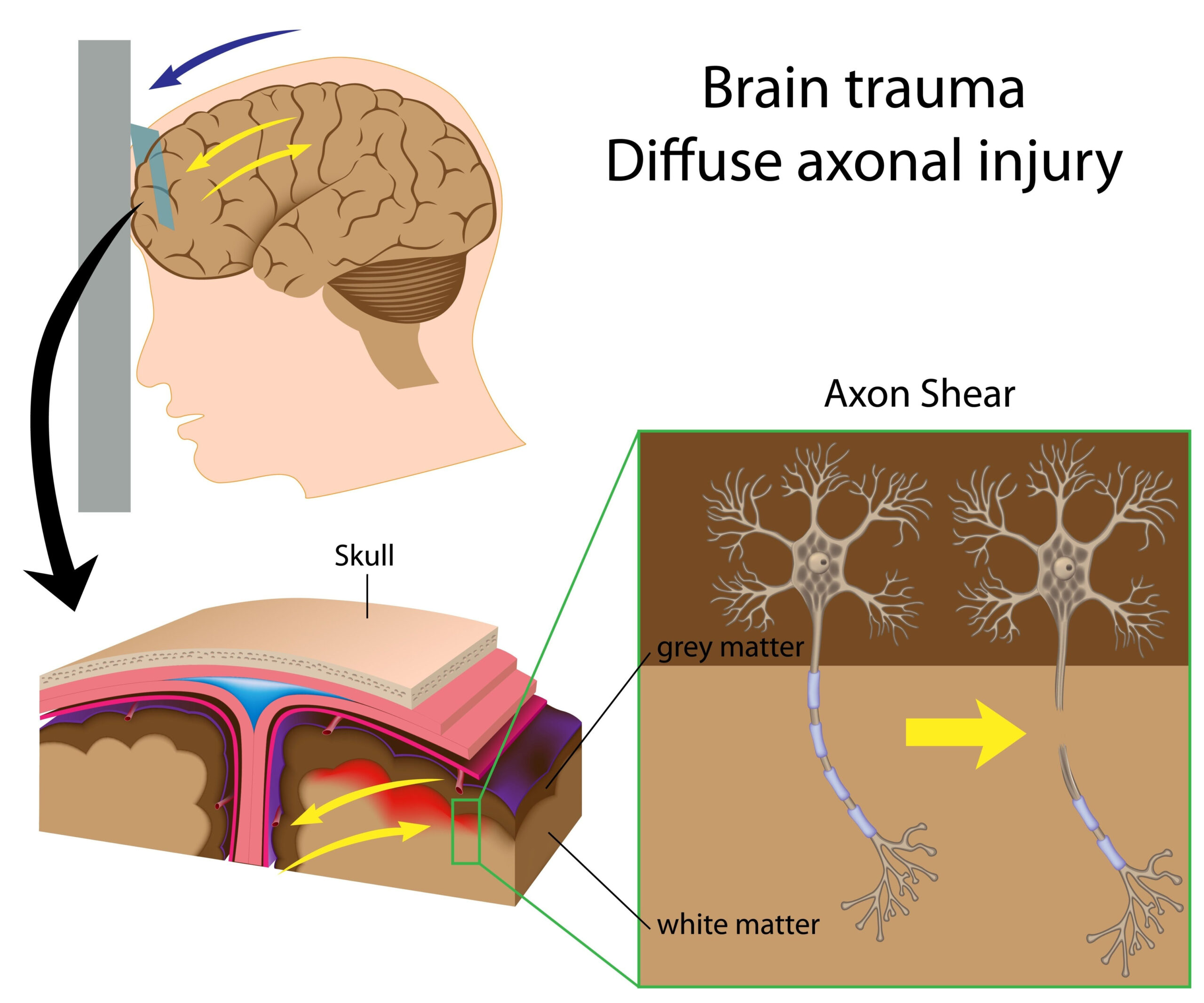
This research focuses on understanding how different kinds of brain connectivity functions in military and civilian individuals suffering from Posttraumatic Stress Disorder (PTSD) and/or Mild Traumatic Brain Injury (MTBI). By examining both dynamic and static resting-state functional connectivity, the study aims to uncover potential differences in brain network interactions among the two populations. Resting-state functional connectivity refers to the shared activity between brain regions when a person is not engaged in a specific task, providing insights into the underlying neural mechanisms associated with these conditions.
The study cohort includes participants diagnosed with PTSD, MTBI, both, or neither condition, allowing for a comparative analysis. This design helps identify unique connectivity patterns in individuals experiencing trauma-related psychological and physical symptoms. By leveraging advanced neuroimaging techniques like functional magnetic resonance imaging (fMRI), researchers are poised to map out the specific brain networks affected by these conditions, contributing valuable knowledge to the field of neuropsychology and potential therapeutic approaches.
The importance of this research lies in its potential to improve our understanding of the neural correlates of PTSD and MTBI, which can lead to more targeted interventions and better treatment outcomes. Identifying specific patterns of brain connectivity may also enhance diagnostic criteria and facilitate the development of personalized medicine approaches for affected individuals.
Methodology
The methodology employed in this study is designed to rigorously assess the differences in brain connectivity between military and civilian populations affected by PTSD and/or MTBI. Participants were carefully selected based on their diagnosis, ensuring a balanced representation from each subgroup: those with PTSD, those with MTBI, individuals diagnosed with both conditions, and a control group without either disorder. This stratified selection allows for a clearer understanding of how each condition manifests at the neurobiological level.
To capture brain activity, the study utilized resting-state functional magnetic resonance imaging (rs-fMRI), a non-invasive imaging technique that measures brain activity by detecting changes associated with blood flow. Participants underwent fMRI scans while at rest, meaning they were instructed to remain still and calm without specific tasks to perform. This resting state is crucial, as it reflects the brain’s intrinsic activity and connectivity networks, offering a window into the underlying mechanisms impacted by PTSD and MTBI.
Dynamic connectivity was analyzed alongside static connectivity. Dynamic connectivity refers to the fluctuations in connectivity patterns over time, highlighting the brain’s adaptability and responsiveness to internal and external stimuli. By using time-varying analysis techniques, the researchers were able to capture transient synchronizations between brain regions, which may be altered in individuals suffering from trauma-related conditions. Conversely, static connectivity provides a comprehensive view of the average connections that remain stable during the resting state, allowing for the identification of enduring neural dysfunctions.
The data analysis incorporated advanced statistical methods to evaluate connectivity patterns. Network-based analyses were employed to identify both overarching and specific alterations in brain function associated with the different diagnoses. This involved implementing machine learning algorithms that can discern patterns not merely through direct observation but by recognizing complex associations and interactions among numerous brain regions.
To ensure the robustness and reproducibility of the findings, the study employed a multi-site approach, gathering data across various research centers. This strategy enhances the generalizability of the results, allowing for a broader understanding of how these neurological changes manifest across diverse populations. Additionally, rigorous protocols were followed for screening participants, maintaining the highest ethical standards, and ensuring that data were collected and processed uniformly across all sites.
Comprehensive cognitive and clinical assessments were conducted alongside neuroimaging to correlate observed brain connectivity patterns with clinical symptoms. These assessments included standardized questionnaires that evaluate PTSD symptoms, cognitive functioning, and other relevant psychological factors. By integrating behavioral data with neuroimaging findings, this research aims to create a more holistic view of how brain connectivity influences the lived experiences of individuals with PTSD and MTBI.
Key Findings
The analysis of brain connectivity patterns revealed several significant differences between military and civilian populations experiencing PTSD and/or MTBI. Notably, distinct alterations in both static and dynamic resting-state connectivity were identified across the various groups, illuminating the complex interplay between these neural changes and the psychological symptoms associated with trauma.
In individuals diagnosed with PTSD, a prominent finding was the disruption in connectivity within the default mode network (DMN), which is primarily active during rest and introspection. The analysis highlighted reduced connectivity between core regions of the DMN, particularly between the medial prefrontal cortex and posterior cingulate cortex, suggesting a potential impairment in self-referential processing and emotional regulation. This disruption aligns with the symptoms of PTSD, where individuals may experience difficulties in reflecting on their thoughts and emotions effectively.
Dynamic connectivity analysis revealed that individuals with PTSD demonstrated more instability in connectivity patterns compared to those without PTSD or MTBI. This instability was characterized by transient connectivity changes that did not facilitate effective communication between brain regions, depending on the demands presented by environmental or internal stimuli. Such findings underscore the possible loss of flexibility in brain networks, a feature that might contribute to heightened anxiety and re-experiencing symptoms characteristic of PTSD.
Among those with MTBI, static connectivity revealed notable alterations in sensorimotor and executive function networks. The compromised integration within these networks implies potential difficulties in maintaining attention and executing tasks, which are common complaints among individuals with MTBI. This finding suggests that the neural basis of cognitive symptoms might differ from the emotional dysregulation observed in PTSD, illuminating the divergent neurobiological underpinnings of these conditions.
Combining results from both dynamic and static analyses also indicated that individuals with both PTSD and MTBI showed the most pronounced connectivity abnormalities across multiple brain networks. Specifically, alterations in the salience network, which plays a crucial role in detecting and filtering relevant stimuli, were particularly evident. This could contribute to heightened sensitivity to stressors and an exaggerated startle response often noted in this population.
The study further examined connectivity patterns in the control group, which exhibited a more stable and integrated pattern of connectivity in comparison to both PTSD and MTBI groups. This stark contrast emphasizes how traumatic experiences can lead to significant disturbances in normal brain functioning, potentially providing biomarkers for identifying at-risk individuals or guiding treatment interventions.
Furthermore, correlations between connectivity alterations and clinical symptom severity were observed. Participants with heightened connectivity disruptions tended to report more severe PTSD symptoms, including intrusive thoughts and emotional numbing. These findings suggest that assessing brain connectivity could not only enhance diagnostic criteria but may also inform treatment strategies, aiming to rectify specific neural disruptions associated with symptom profiles.
The findings support the hypothesis that PTSD and MTBI are characterized by distinct yet overlapping patterns of brain connectivity alterations. This nuanced understanding highlights the importance of considering these conditions not only as co-occurring disorders but as unique manifestations with differing neurobiological signatures. The implications of these insights extend toward the development of targeted therapies aimed at restoring connectivity within implicated brain networks, potentially improving therapeutic outcomes for affected individuals.
Clinical Implications
The insights gathered from this study hold significant clinical implications for the diagnosis and treatment of individuals affected by PTSD and/or MTBI. Understanding the specific patterns of brain connectivity alterations offers a foundation for developing targeted therapeutic interventions. The disruptions observed in both static and dynamic connectivity may serve as biomarkers for these conditions, aiding clinicians in early identification and tailoring treatment plans to individual needs based on their unique neurobiological profiles.
For instance, the identification of impaired connectivity within the default mode network in PTSD patients may prompt the use of cognitive-behavioral therapies that specifically aim to improve self-referential processing and emotional regulation. Therapeutic approaches, such as mindfulness and other introspective techniques, could be integrated to enhance connectivity in affected regions, thereby potentially alleviating symptoms related to emotional dysregulation. Moreover, these target-specific strategies could be adapted based on whether a patient exhibits more dynamic instability or static connectivity alterations.
In the case of individuals with MTBI, the alterations detected in sensorimotor and executive function networks suggest that rehabilitation strategies may need to focus on cognitive training and exercises that reinforce attention and task execution. Personalized cognitive rehabilitation could be structured to strengthen the affected brain networks, thereby helping enhance functional outcomes. The consideration of brain connectivity profiles when implementing such therapies could allow for a more effective recovery by addressing the precise neural disruptors at play.
Moreover, the findings underscore the relevance of comprehensive clinical assessments that integrate neuroimaging data with behavioral symptoms. Clinicians can utilize this combined approach not only to enhance diagnostic accuracy but also to monitor treatment effectiveness over time. By observing changes in brain connectivity alongside symptom relief, healthcare providers can make informed decisions regarding the continuation, modification, or escalation of therapeutic interventions.
Importantly, establishing these brain connectivity patterns in connection with symptom severity manifestations suggests that researchers and clinicians alike should prioritize investigating connectivity metrics as potential endpoints in clinical trials. This shift could lead to novel treatment approaches that are both innovative and deeply rooted in the underlying neural mechanisms tied to PTSD and MTBI.
These findings illuminate the complexity of brain connectivity in trauma-related conditions and emphasize the need for a nuanced understanding of each disorder. By shifting clinical practices to incorporate neurobiological insights gained from research, there exists great potential for enhancing therapeutic outcomes and improving quality of life for individuals grappling with the lasting effects of trauma.


