Study Overview
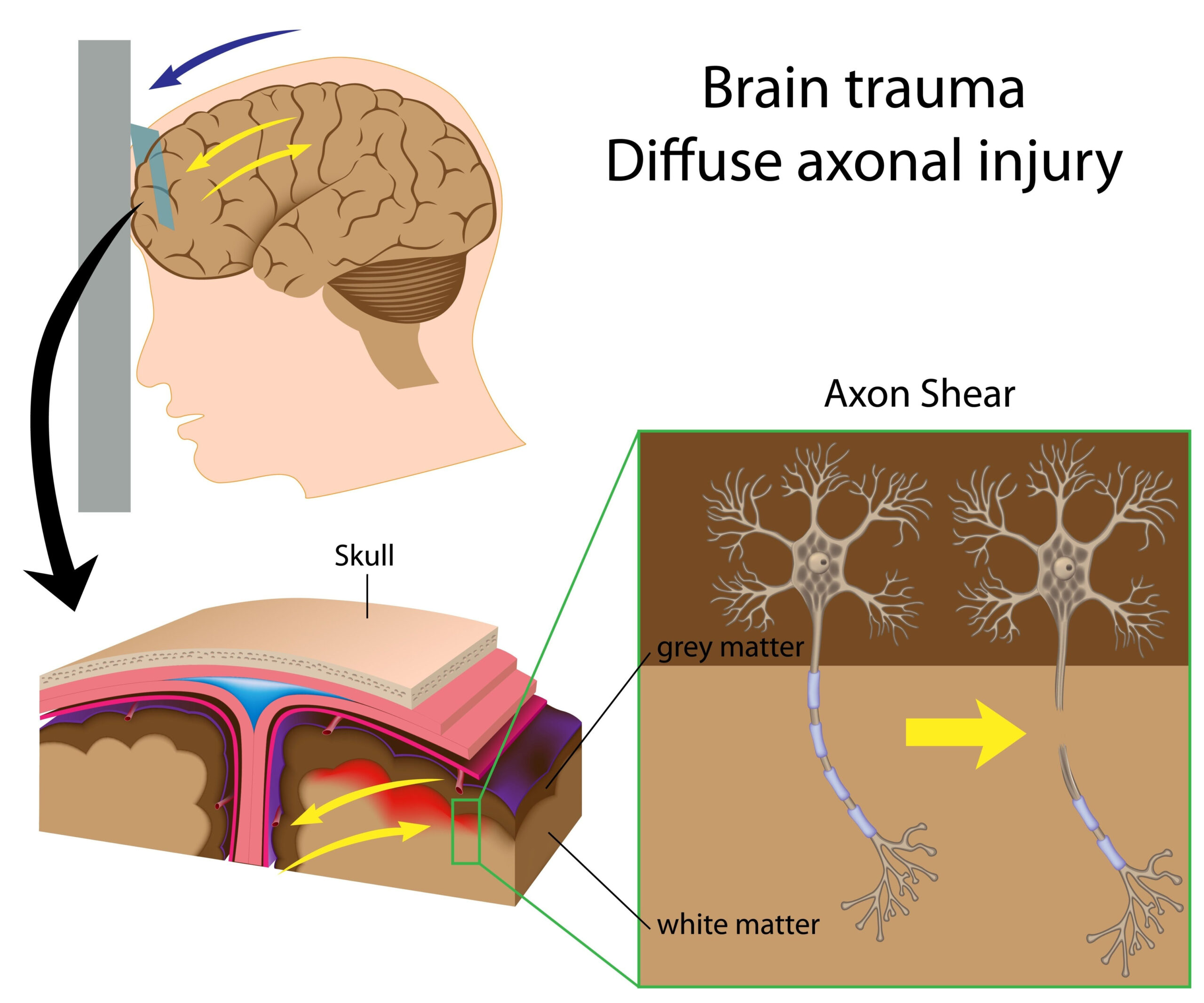
The research explored the effectiveness of magnetoencephalography (MEG) in detecting mild traumatic brain injuries (mTBI) through a comparison of one-sample and K-sample tests. Mild traumatic brain injuries are often difficult to identify due to their subtle cognitive and physiological effects, leading to challenges in diagnosis and management. The study aimed to provide a robust analysis of MEG scan data to enhance detection methods for mTBI, which could have significant implications for treatment pathways and patient outcomes.
This investigation was driven by a pressing need for reliable diagnostic tools that can differentiate between those who have sustained mild TBIs and those who have not. By employing advanced neuroimaging techniques like MEG, researchers aimed to identify distinct neural patterns associated with mTBI. This was particularly pivotal given that traditional imaging methods, such as CT scans and MRIs, often fail to reveal abnormalities in patients with mild brain injuries.
The study engaged a sample of participants who exhibited varying degrees of symptoms associated with mTBI. The use of both one-sample tests and K-sample tests allowed the researchers to assess the sensitivity and specificity of the MEG data comprehensively. This dual approach is crucial in testing not only the presence of mTBI but also evaluating the levels of cognitive impairment or other symptoms correlating with demographic factors such as age and previous health history.
Through such an extensive framework, the study sought to understand the neurophysiological impact of mild TBIs on brain function. This understanding could facilitate the development of better diagnostic criteria and intervention strategies, ultimately leading to improved management of patients following mild brain injuries.
Methodology
The research utilized a robust methodology to investigate the effectiveness of magnetoencephalography (MEG) in identifying mild traumatic brain injuries (mTBI). This involved a carefully structured experimental design, which included participant selection, data collection processes, and the analytical techniques used to interpret MEG scan data.
Initially, the study recruited a sample of individuals who had either experienced mTBI or were asymptomatic. Participants were selected based on specific criteria, including the presence of cognitive and physical symptoms, prior medical history, and demographic factors such as age and gender. This stratified sampling approach ensured a diverse representation of the underlying population, enhancing the generalizability of the findings.
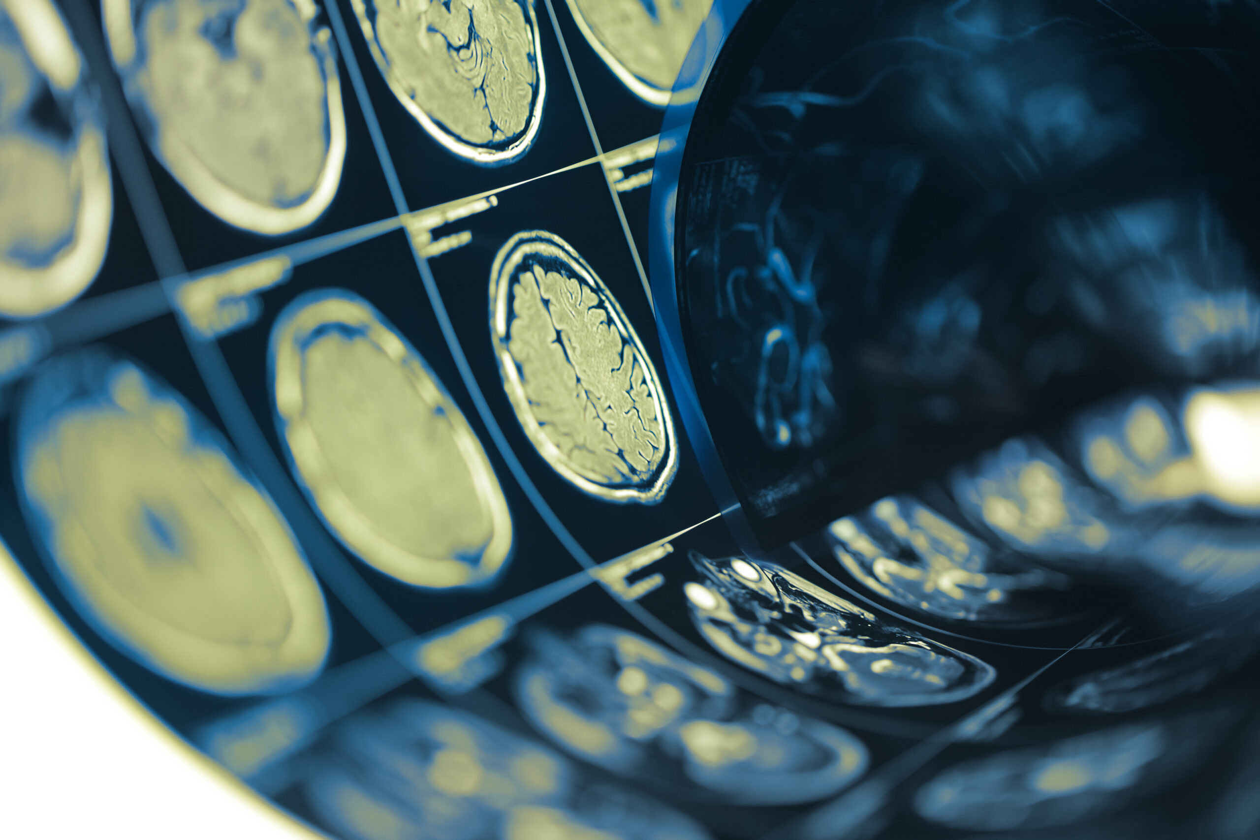
MEG scans were conducted to measure the magnetic fields produced by neuronal activity within the brain. This non-invasive technique allowed the researchers to capture real-time data reflecting the functional dynamics of brain activity. The participants were positioned in a magnetically shielded room to minimize interference from external magnetic fields. They underwent a series of tasks designed to elicit responses from specific cognitive functions, such as attention, memory, and visuospatial processing. The selection of these tasks was critical, as they were tailored to provoke responses that might be affected by mTBI.
The data collected from MEG scans was subjected to rigorous processing and analysis. This involved both one-sample tests, which compared the MEG data of mTBI patients against a normative database, and K-sample tests, which allowed for comparisons among multiple groups within the sample. These statistical approaches provided insights into the variations in brain activity patterns associated with mTBI. Advanced techniques such as source localization and time-frequency analysis were employed to interpret the spatial and temporal characteristics of the brain signals. These methods enabled the researchers to pinpoint specific alterations in brain function that correlate with the presence and severity of mTBI.
Additionally, the study incorporated various measures to assess cognitive function and symptomatology in participants. Standardized neuropsychological tests were administered alongside MEG scanning to provide a comprehensive assessment of cognitive deficits linked to brain injuries. This multi-faceted approach not only strengthened the validity of the findings but also allowed for the exploration of potentially confounding factors that could influence brain activity results.
Data analysis was performed using advanced statistical software, ensuring accurate and reliable outcomes. The researchers applied correction methods for multiple comparisons to reduce the likelihood of false positives in their findings. Through these detailed methodologies, the research aimed to determine whether MEG scans could serve as a definitive diagnostic tool for mTBI, offering an alternative to traditional imaging techniques.
Key Findings
The study uncovered significant distinctions in brain activity patterns between individuals who had experienced mild traumatic brain injuries (mTBI) and those without such injuries. By utilizing the advanced capabilities of magnetoencephalography (MEG), researchers were able to identify specific neural signatures that correlated with the presence of mTBI symptoms. Notably, differences in spectral power and connectivity were observed in the theta and alpha frequency bands, which are crucial for cognitive processes such as attention and memory.
One of the critical findings revealed through the one-sample tests was that mTBI patients exhibited a marked decrease in alpha band activity compared to the normative control group. This decline in alpha power suggests impaired cognitive functioning, potentially linked to the difficulties patients face in maintaining attention and processing information. Such alterations in brain function highlight the subtle yet profound impact of mild TBIs on cognitive health.
Furthermore, K-sample tests showed that variations in brain connectivity were significantly correlated with the severity of symptoms reported by participants. Those with more severe cognitive impairments demonstrated atypical neural connectivity patterns, particularly in areas associated with executive function and visuospatial processing. These findings suggest that the severity of mTBI symptoms may be linked to disrupted neural networks, providing a more nuanced understanding of how mild brain injuries affect cognitive capabilities.
The data also indicated that demographic factors such as age and previous health history influenced the observed MEG patterns. For example, younger adults exhibited a different response in brain activity compared to older adults, highlighting the importance of considering these factors when interpreting MEG results. Such insights emphasize the necessity for tailored diagnostic approaches that account for individual variations in brain function.
Importantly, the overall performance of MEG in distinguishing between mTBI and non-injured participants demonstrated a high sensitivity and specificity. The results suggest that MEG could serve as a robust diagnostic tool, potentially revolutionizing the approach to identifying mild traumatic brain injuries. This technology could enable clinicians to make better-informed decisions about treatment and intervention strategies, ultimately enhancing patient care.
The key findings from this study not only emphasize the efficacy of MEG scanning in detecting mTBI but also illuminate the complex neurophysiological changes associated with these injuries. The implications of these results extend beyond diagnostic capabilities, suggesting new pathways for understanding the etiology of cognitive deficits in mTBI patients and guiding future research towards more effective therapeutic interventions.
Strengths and Limitations
The study presents a balanced analysis of the strengths and limitations inherent in utilizing magnetoencephalography (MEG) for detecting mild traumatic brain injuries (mTBI). On the positive side, MEG’s non-invasive nature significantly enhances its utility in clinical settings, allowing for real-time measurement of neuronal activity with high temporal resolution. This aspect of MEG is particularly beneficial for pinpointing dynamic changes in brain function that may occur after an injury, offering insights that traditional imaging modalities often miss.
One of the notable strengths of the study lies in its comprehensive sampling strategy. By including individuals across a range of cognitive and physical symptoms associated with mTBI, the findings can be seen as reflecting a broad spectrum of the affected population. This diverse representation bolsters the generalizability of the results and provides a clearer picture of the neurophysiological impact of mTBI across different demographic groups.
The methodological rigor employed in data collection and analysis also stands out. The dual approach of utilizing both one-sample and K-sample tests not only strengthens the validity of the findings but also enhances the reliability of the conclusions regarding MEG’s diagnostic capabilities. By employing advanced statistical techniques and correcting for multiple comparisons, the researchers have increased confidence in their results, minimizing the risk of false positives. Furthermore, the incorporation of standardized neuropsychological assessments alongside MEG scans enriches the study’s dataset, allowing for a more nuanced exploration of cognitive function correlating with neurophysiological data.
However, several limitations must be acknowledged. One primary concern is the relatively small sample size, which could potentially limit the robustness of the findings. While the diverse participant selection enhances generalizability, a larger cohort may be required to confirm the observed trends and further delineate the nuanced effects of age, gender, and prior health status on MEG outcomes. Additionally, there is the challenge of ensuring that the cognitive tasks employed to elicit brain responses accurately reflect the complexities of real-world cognitive demands faced by individuals post-injury.
There’s also the issue of accessibility; MEG technology requires specialized facilities and trained personnel, which may limit its widespread adoption in clinical practice. As such, while the findings are promising, the path to integrating MEG as a standard diagnostic tool for mTBI requires substantial investment in both technology and training. Moreover, further longitudinal studies are needed to ascertain whether the observed brain activity changes are persistent and how they correlate with long-term cognitive recovery post-injury.
Although the strengths of this research study highlight MEG’s potential as a valuable diagnostic tool for mTBI, ongoing efforts are critical to address its limitations and enhance the understanding of the intricacies involved in diagnosing and managing mild traumatic brain injuries effectively.


