Neurobiological Mechanisms
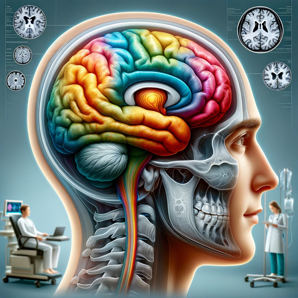
Understanding the neurobiological mechanisms underlying altered saccades, blink rates, and pupil responses in functional motor disorders is essential for grasping how these phenomena are linked to the brain’s functionality and overall motor control. Recent studies have suggested that these involuntary eye movements are not merely peripheral manifestations but rather indicative of a more complex interplay within the central nervous system.
Research indicates that the areas of the brain responsible for saccadic eye movements—primarily the frontal eye fields (FEF), supplementary eye fields (SEF), and the superior colliculus—may exhibit altered activity patterns in individuals with functional motor disorders. These regions are crucial for initiating rapid eye movements and coordinating visual attention, thus their dysfunction can lead to significant impairments in voluntary motor control and eye movement precision.
Furthermore, neuroimaging studies have highlighted abnormalities in connectivity between these areas and other related regions, including the basal ganglia and the cerebellum. These connections are integral for integrating sensory information and executing smooth motor responses. As such, disruptions in this circuitry may contribute to the characteristic presentation of saccadic dysregulation seen in patients.
In addition to altered saccadic movements, pupillary responses also offer insight into the neurobiological mechanisms at play. The pupil dilation reflex, which is mediated by the autonomic nervous system, can be affected by emotional and cognitive states. In individuals with functional motor disorders, variations in pupil size under differing stimulus conditions could point to dysregulation in the brain’s emotional and arousal pathways, particularly those involving the locus coeruleus and the autonomic nervous system.
One of the most compelling findings involves the role of cognitive processes in these motor control functions. Unlike traditional neurological disorders that exhibit clear-cut physical damage, functional motor disorders often entail a complex relationship between cognitive expectations and motor output. Patients might exhibit normal reflexes under certain conditions or when attention is diverted, suggesting that the underlying issue may involve higher-order cognitive control rather than solely peripheral dysfunction.
| Neurobiological Area | Function | Alterations in Functional Motor Disorders |
|---|---|---|
| Frontal Eye Fields (FEF) | Control of voluntary eye movements | Altered activation patterns leading to saccadic dysregulation |
| Supplementary Eye Fields (SEF) | Preparation and execution of eye movements | Potential disruptions in motor planning processes |
| Superior Colliculus | Integration of sensory input and eye movement coordination | Impaired coordination between sensory and motor systems |
| Locus Coeruleus | Pupil dilation and arousal | Dysregulation in autonomic functioning affecting pupil responses |
These insights emphasize a multi-faceted understanding of the neurobiological underpinnings of altered saccades, blink, and pupil responses in functional motor disorders. Recognizing that the root causes may extend beyond conventional neurological impairments opens new avenues for investigation and potential therapeutic strategies, particularly those targeting the cognitive and emotional components of motor control.
Clinical Presentations
Physiological Assessments
To effectively evaluate the alterations in saccades, blink rates, and pupil responses associated with functional motor disorders, a variety of physiological assessments have been employed. These assessments play a critical role in understanding the interplay between neurobiological mechanisms and clinical presentations. They help elucidate the response patterns observed in patients and provide valuable insight into the dysregulation of motor systems.
One of the primary methods used to assess these involuntary eye movements is saccadic eye movement analysis. This involves monitoring the speed, amplitude, and frequency of saccadic movements under various visual stimuli. Research shows that individuals with functional motor disorders often display slower saccadic velocities and increased latency, suggesting a fundamental disruption in the neural processes that facilitate rapid eye movements. In expansive studies using high-speed eye trackers, variations in these parameters can be quantitatively assessed and compared against healthy controls, providing a clearer picture of the motor control deficits present in affected individuals.
Furthermore, blink rate assessments allow researchers to gauge the efficiency of autonomic control. Typically, blink rates are influenced by environmental stimuli, cognitive load, and emotional state. In functional motor disorders, abnormal blink rates—either excessively high or low—can indicate maladaptive responses involving the brain’s emotional processing networks. For example, studies have observed that individuals under increased cognitive stress may exhibit reduced blink rates, a phenomenon attributed to heightened focus or arousal state that overshadows basic reflexive actions.
Pupil response analysis is another vital physiological assessment that provides insights into autonomic dysfunction. Pupil size and reactivity are influenced by both cognitive engagement and emotional states. In tests of pupil reactivity, patients with functional motor disorders frequently exhibit abnormal dilation responses to light changes or emotional stimuli. This deviation suggests disruptions in the pathways connecting the locus coeruleus and the pupillary control centers, correlating pupil dynamics with cognitive and emotional processes.
Table 1 summarizes the physiological assessments relevant to understanding the alterations in functional motor disorders:
| Assessment Type | Measurement Aspect | Expected Findings in Functional Motor Disorders |
|---|---|---|
| Saccadic Eye Movement Analysis | Speed, amplitude, frequency of saccades | Slower velocities, increased latency of saccadic movements |
| Blink Rate Assessment | Frequency of blinks under varied conditions | Abnormal blink rates (either high or low) indicating autonomic dysregulation |
| Pupil Response Analysis | Pupil size and reactivity to light and emotional stimuli | Altered reactivity patterns suggesting disruptions in autonomic control mechanisms |
These physiological assessments not only help in diagnosing functional motor disorders but also assist in mapping the neurobiological landscape influencing eye movements and autonomic responses. As research progresses, these assessments may uncover new avenues for targeted interventions aimed at restoring normal functional capabilities in affected individuals.
Future Research Directions
Future research exploring the neurobiological underpinnings of functional motor disorders must focus on several key areas to enhance our understanding and treatment of altered saccades, blink rates, and pupil responses. One promising avenue involves the integration of advanced neuroimaging techniques, such as functional magnetic resonance imaging (fMRI) and magnetoencephalography (MEG), to observe real-time brain activity during specific tasks that engage eye movement and autonomic responses. This approach may reveal how disruptions in brain networks correlate with the clinical manifestations of these disorders. By identifying unique biomarkers associated with altered neural connectivity, clinicians could potentially develop more tailored therapeutic strategies.
Furthermore, longitudinal studies are essential for understanding the trajectory of functional motor disorders over time. Tracking changes in eye movement patterns and autonomic responses among individuals with these disorders could offer insights into progression and recovery. Such studies would also allow for the assessment of treatment efficacy, especially when comparing behavioral interventions, cognitive therapy techniques, or pharmacological treatments aimed at restoring normal autonomic functions.
Another critical area for exploration lies in the cognitive factors contributing to functional motor disorders. Investigating how elements such as anxiety, depression, and expectation influence motor control could lead to the development of integrated therapeutic approaches targeting both psychological and physiological aspects of the disorder. Experimental designs that manipulate cognitive load or emotional stimuli can further clarify these relationships by observing corresponding changes in saccadic and pupillary responses.
Additionally, expanding research to include diverse populations may enhance the generalizability of findings. Variability in genetic, cultural, and socio-economic backgrounds can impact the expression and experience of functional motor disorders. Investigating these differences could reveal specific risk factors or protective mechanisms relevant to particular groups, guiding more effective interventions.
Finally, the introduction of artificial intelligence and machine learning in analyzing data from physiological assessments may revolutionize how we interpret the complex interplay between neurobiological mechanisms and clinical symptoms. Advanced algorithms can uncover patterns that are not readily visible to human observers, aiding in diagnostic precision and prognostic evaluations.
Table 1 summarizes potential future research directions that could further elucidate the mechanisms behind functional motor disorders:
| Research Direction | Focus Area | Potential Impact |
|---|---|---|
| Neuroimaging Studies | Real-time brain activity during eye movement tasks | Identifying biomarkers for personalized treatments |
| Longitudinal Studies | Tracking changes over time in individuals | Understanding disorder progression and treatment efficacy |
| Cognitive Contributions | Influence of psychological factors on motor control | Developing integrated therapeutic approaches |
| Diverse Population Studies | Exploring variations across different demographics | Identifying risk factors and tailored interventions |
| AI and Machine Learning | Analyzing complex physiological data | Enhancing diagnostic precision and prognostic abilities |
By pursuing these research avenues, the field can gain a more comprehensive understanding of the mechanisms underlying functional motor disorders and work towards innovative solutions that improve patient outcomes.