Study Overview
The investigation centers on the relationship between blood-based biomarkers and neuroimaging outcomes in individuals experiencing mild traumatic brain injury (mTBI). mTBI is characterized by transient neurological dysfunction resulting from mechanical forces to the head, and it frequently manifests in sports and accidents. Understanding how blood biomarkers correlate with neuroimaging results could potentially enhance diagnostic accuracy and treatment protocols for mTBI.
This study involved a thorough analysis of patients who presented with mTBI symptoms and underwent both blood tests and neuroimaging scans. The primary objective was to identify specific biomarkers in the bloodstream that could be linked to observable changes in brain imaging, particularly utilizing techniques such as MRI and CT scans. Researchers hypothesized that certain protein levels in the blood, indicative of neuronal damage or inflammation, would correlate with specific patterns or abnormalities observed in imaging studies.
The cohort consisted of participants with varied clinical presentations, allowing for a comprehensive exploration of the biomarkers’ applicability across a range of mTBI cases. The study systematically assessed blood samples for commonly recognized biomarkers, including S100B, GFAP, and UCH-L1, which are known for their association with brain injury. Simultaneously, advanced imaging techniques were employed to visualize brain structure and identify any underlying trauma or morphological changes resulting from the injury.
By employing both quantitative biomarker assessment and qualitative neuroimaging analysis, the study aimed to uncover significant correlations that could inform clinical practice. The findings are anticipated to advance understanding of mTBI and potentially lead to the development of standardized diagnostic tools that incorporate both blood tests and imaging assessments to improve patient outcomes.
Biomarker Analysis
Within this study, the focus was placed on several key blood-based biomarkers known to reflect neuronal damage and inflammation, assessed in patients with mild traumatic brain injury (mTBI). The biomarkers selected, S100B, GFAP (Glial Fibrillary Acidic Protein), and UCH-L1 (Ubiquitin C-terminal hydrolase L1), are integral to understanding the pathophysiological processes that occur following a traumatic event in the brain.
S100B is a calcium-binding protein predominantly found in astrocytes, which are crucial for maintaining the brain’s homeostasis. Elevated levels of S100B in circulation have been linked to cerebral damage and poor outcomes following various types of brain injury. Its significance lies in its potential as a non-invasive marker of neuronal injury, allowing clinicians to gauge the extent of damage without requiring invasive procedures.
GFAP serves as an important biomarker for astroglial activation, and its presence in the bloodstream can indicate neuroinflammation and cellular stress following trauma. Increased GFAP levels post-injury correlate with the severity of the brain injury and have been shown to be predictive of long-term cognitive outcomes. This makes GFAP a valuable tool for assessing the impact of mTBI on brain health and recovery trajectories.
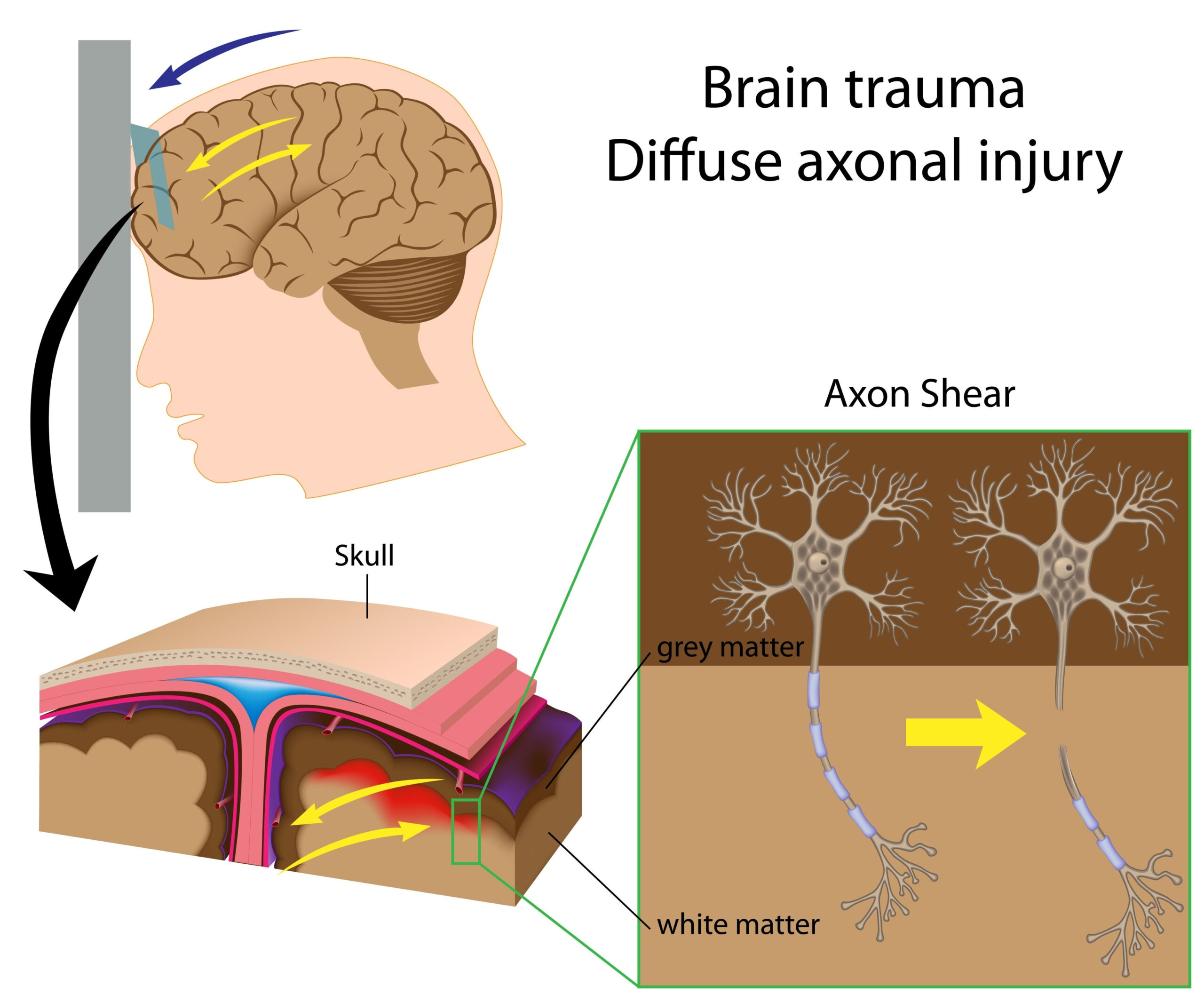
UCH-L1 is another promising biomarker, reflecting axonal injury and cell degradation. Typically found in neurons, its release into the bloodstream suggests significant neuronal damage. Studies have shown that UCH-L1 levels can provide insights into the extent of axonal injury, making it a critical component in the assessment of mTBI.
The analysis conducted in this study involved measuring these biomarkers in blood samples collected from participants with confirmed mTBI. The results were then compared to neuroimaging findings utilized for diagnostic purposes, with the aim of identifying correlations between specific biomarker levels and the characteristics of the imaging results. Such correlations are essential not only for confirming the presence of brain injury but also for monitoring recovery and guiding treatment strategies. In essence, this dual approach of analyzing blood-based biomarkers alongside neuroimaging data holds potential for stratifying patients based on injury severity and tailoring management plans accordingly.
Emerging data from the study underscored the complex interplay between these biomarkers and neuroimaging findings, revealing that elevated levels of specific biomarkers were often observed in conjunction with distinct neuroimaging abnormalities. For example, patients exhibiting higher S100B levels frequently presented with noticeable edema on MRI scans, suggesting a direct relationship between biomarker release and observable brain alterations.
Furthermore, the study aimed to provide clarity on the temporal dynamics of these biomarkers in relation to symptomatology and imaging changes. For example, understanding how these biomarkers fluctuate over time post-injury could be pivotal in determining not just initial injury severity but also in predicting recovery patterns. This has profound implications for clinical practice, where timely decision-making can be crucial to effective patient care.
Ultimately, the insights gained from the biomarker analysis may serve to not only inform diagnostic strategies but also encourage a more personalized approach to treatment, where interventions are tailored based on both biomarker levels and the severity of imaging findings. Addressing the urgent need for refined diagnostic tools in mTBI management could markedly improve outcomes and quality of care for affected individuals.
Imaging Techniques
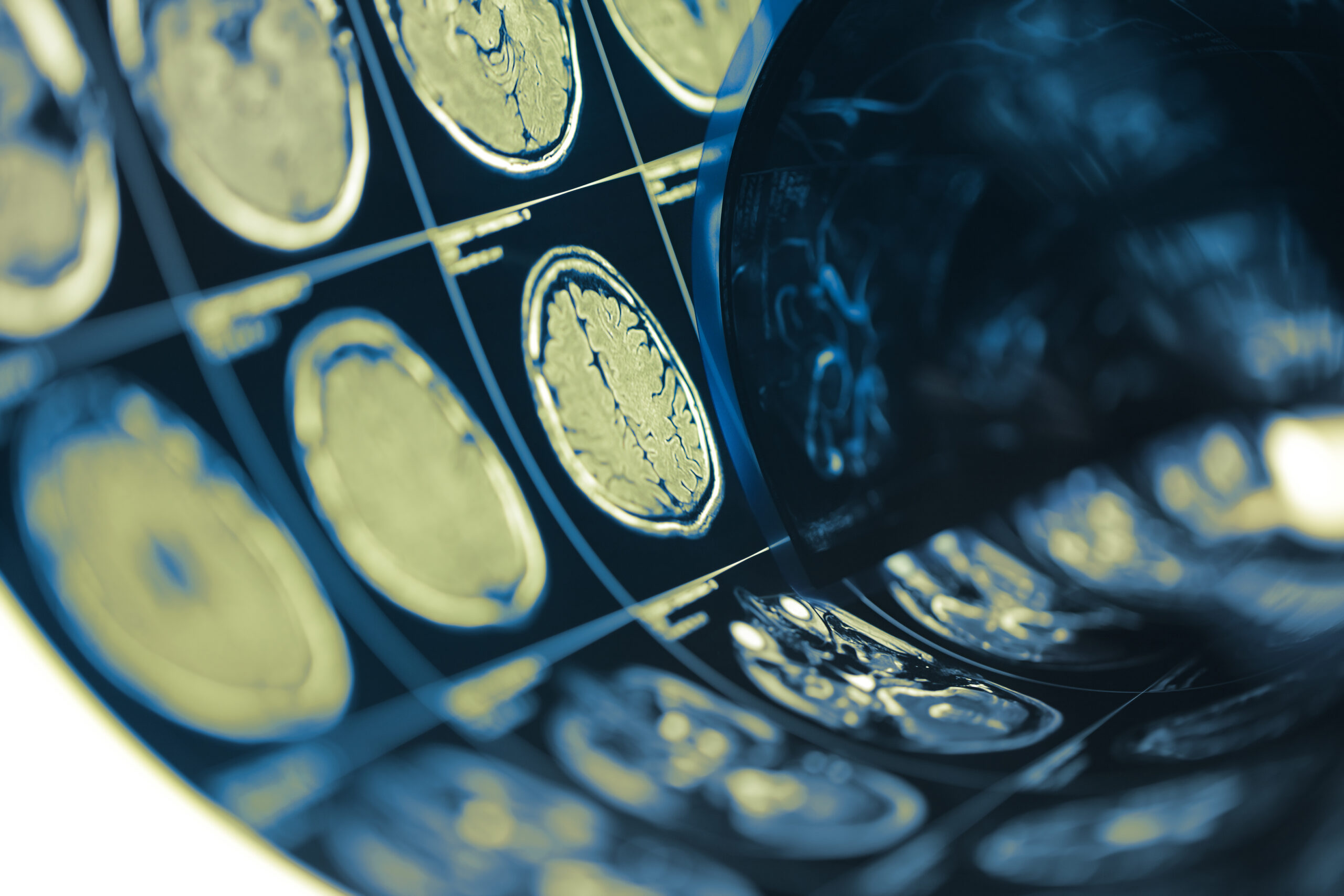
The study utilized advanced neuroimaging techniques to assess the structural and functional abnormalities in the brains of participants with mild traumatic brain injury (mTBI). Magnetic resonance imaging (MRI) and computed tomography (CT) emerged as the primary modalities for this investigation, each offering unique advantages in visualizing brain injuries and understanding the underlying pathology of mTBI.
MRI is particularly well-suited for detailing soft tissue structures, providing high-resolution images that can reveal subtle changes in brain morphology. This technique employs powerful magnetic fields and radio waves to generate detailed images, making it invaluable for identifying issues such as edema, hemorrhages, and axonal injuries that may not be readily apparent on other imaging modalities. In the context of mTBI, MRI can reveal microstructural changes linked to neuronal integrity and connectivity, thereby enhancing understanding of the injury’s impact on brain function.
Meanwhile, CT scanning is often the first-line imaging technique in acute trauma settings. It is rapid and highly effective in detecting acute intracranial hemorrhages and skull fractures. In this study, CT scans provided an immediate assessment of potential life-threatening injuries and were essential in the initial evaluation of patients presenting with symptoms of mTBI. However, while CT is adept at identifying gross structural anomalies, it may overlook subtle brain changes compared to MRI.
The integration of these imaging techniques allowed for a comprehensive evaluation of brain function and structure post-injury. Participants underwent both MRI and CT scans, enabling researchers to correlate imaging findings with blood biomarker levels. This approach provided insights into how specific neuroimaging features corresponded to the biochemical evidence of neuronal damage or inflammation reflected in the blood biomarkers.
For instance, standardized imaging protocols enabled the classification of neuroimaging abnormalities based on severity and location. The study particularly noted the presence of diffuse axonal injury on MRI, with corresponding elevations in biomarkers such as UCH-L1 suggesting a strong linkage between clinical signs of mTBI, biochemical markers, and observable imaging defects. Additionally, the analysis explored the occurrence of localized edema or ischemic changes noted on MRI scans in conjunction with heightened concentrations of S100B, reinforcing the hypothesis that neuroimaging findings can elucidate the extent of neurobiological disruption following mTBI.
Moreover, the research delved into the timing of imaging assessments in relation to symptom manifestation and biomarker levels. Identifying the optimal window for imaging—both in terms of initial evaluation and follow-up studies—was crucial for understanding the evolution of mTBI. This temporal analysis not only aided in correlating symptom severity with structural changes but also illuminated pathways to optimize patient management and recovery tracking.
Through these comprehensive imaging strategies, the study highlighted the potential of integrating neuroimaging findings with blood biomarkers to enhance diagnostic precision. The combined analysis may pave the way for developing an evidence-based approach to stratify patients based on the severity of their mTBI, facilitating targeted therapeutic interventions. As our understanding deepens through these methods, the goal remains to refine clinical pathways, improve patient outcomes, and foster a more nuanced appreciation of the dynamics of mTBI.
Implications for Treatment
The implications of this study extend significantly into the treatment landscape for mild traumatic brain injury (mTBI). By elucidating the connections between blood-based biomarkers and neuroimaging findings, clinicians may be better equipped to tailor treatment strategies to individual patient profiles, leading to more effective management of mTBI over time.
A clear outcome of the biomarker analysis indicates that specific biomarkers correlate with the severity of brain injury as observed through imaging techniques. For instance, patients with elevated levels of S100B not only showed imaging evidence of edema but also reported more severe symptomatic presentations. This correlation suggests that clinicians could employ biomarker testing as a predictive tool to determine the potential severity of injury and, consequently, the urgency and intensity of treatment interventions.
Adopting a biomarker-informed treatment strategy could facilitate a shift from a one-size-fits-all approach to a more personalized model of care. With evidence indicating that GFAP levels correlate with the timeline of recovery, clinicians may decide to adjust rehabilitation protocols based on real-time biomarker feedback. For example, patients exhibiting prolonged high levels of GFAP might benefit from more aggressive therapeutic measures, such as cognitive rehabilitation and targeted physical therapies, aimed at promoting recovery and mitigating long-term effects.
Furthermore, integrating blood biomarkers with neuroimaging outcomes enhances the precision of diagnosing complications associated with mTBI. Individuals who present with elevated UCH-L1 levels alongside specific neuroimaging findings may require closer monitoring for secondary injuries or complications. This proactive stance could allow for timely intervention, potentially preventing worsening outcomes and facilitating a smoother recovery journey.
The potential for early identification of recovery trajectories, as indicated by biomarker fluctuations, can also support decisions regarding return-to-play or return-to-work protocols. If certain biomarkers signal a delayed recovery, clinicians might recommend extended observation periods, ensuring that patients do not prematurely resume activities that could exacerbate their conditions.
Moreover, the research sets the stage for further investigations into targeted therapies. Given the insights provided by the biomarkers regarding different injury pathways within the brain, there may be an opportunity to explore pharmacological or non-pharmacological interventions that specifically address the underlying mechanisms of neuronal damage and neuroinflammation. Interventions could be designed based on individualized biomarker profiles, optimizing therapeutic efficacy and promoting brain health recovery.
While the findings present a promising direction for clinical practice, it is also important to underscore the need for continuous research and refinement of treatment guidelines based on evolving biomarker science. Establishing standardized protocols for integrating biomarker assessments and imaging findings into clinical workflows will be paramount for ensuring that the potential benefits translate into improved patient outcomes on a broader scale.
The convergence of blood biomarker analysis with neuroimaging outcomes not only validates the physiological underpinnings of mTBI but also reinforces the necessity for interdisciplinary collaboration among clinicians, researchers, and rehabilitation specialists. The implications are profound, with the potential to reshape the landscape of mTBI treatment by emphasizing personalized, data-driven, and timely therapeutic interventions.