Study Overview
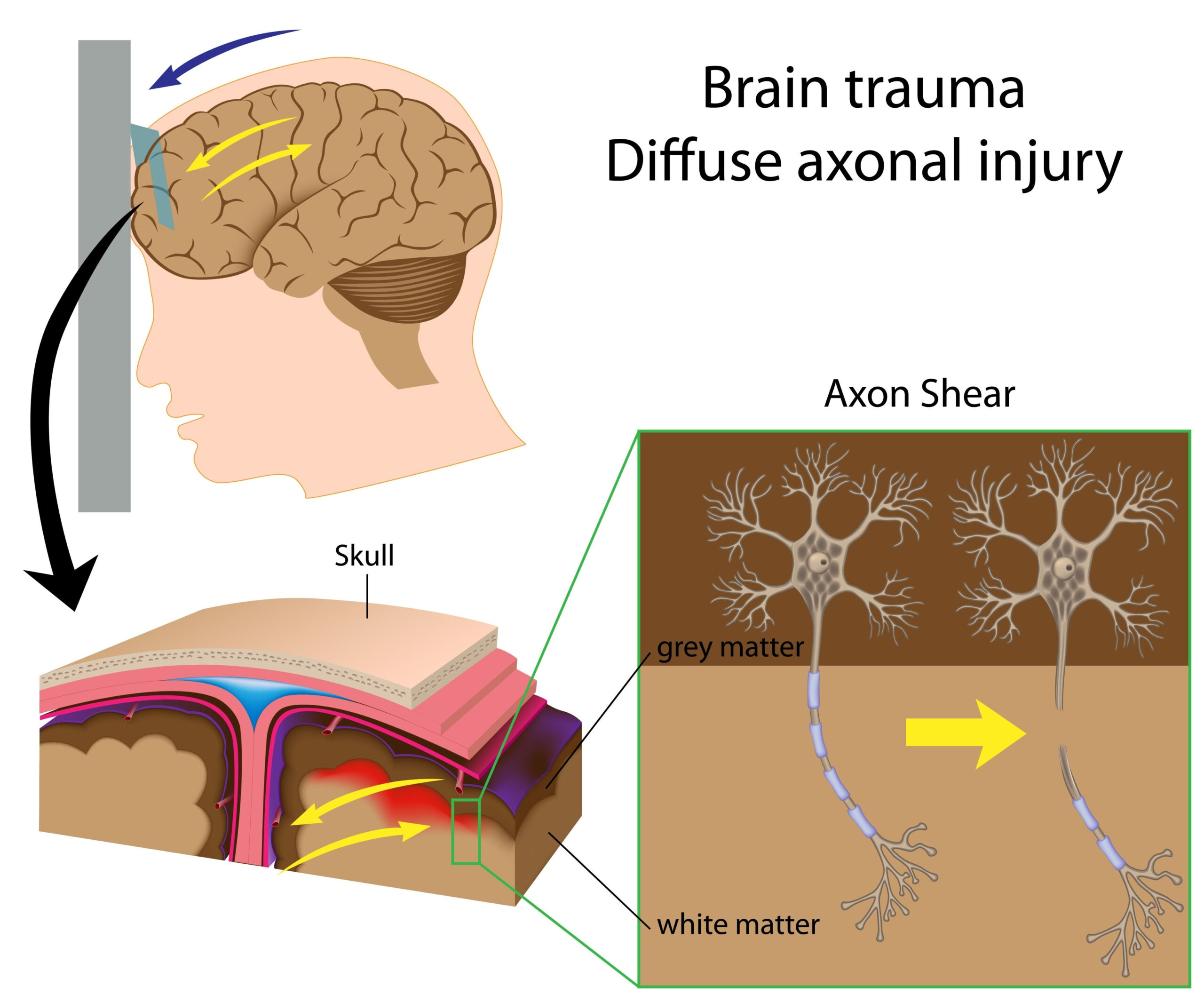
The research focuses on the innovative use of magnetic particle imaging (MPI) as a diagnostic tool for detecting traumatic brain injury (TBI) in a living organism. Traumatic brain injuries are complex conditions that arise from external forces impacting the head, potentially leading to significant, long-lasting neurological consequences. Traditional imaging techniques, such as CT and MRI, offer valuable insights but often fall short in sensitive detection of small injury foci.
In this study, the researchers sought to explore how MPI, which utilizes superparamagnetic nanoparticles, can provide enhanced visualization of brain injuries. MPI operates by detecting the magnetic signals emitted from these nanoparticles, allowing for real-time imaging with high spatial resolution and minimal background noise. This method not only promises to improve the identification of minor brain injuries but also stands to enhance the understanding of biochemical processes associated with TBIs.
The investigation involved a systematic approach, where experimental models were subjected to controlled traumatic events. Following these events, MPI was employed to visualize potential injuries. The researchers meticulously recorded imaging results alongside conventional diagnostic methods to establish a comparative framework. The key objective was to evaluate the efficacy of MPI in not only identifying injury locations but also gauging the extent of the tissue damage.
Overall, this study represents a stride toward integrating advanced imaging technologies in clinical settings, ultimately aiming to refine diagnosis and treatment strategies for individuals suffering from traumatic brain injuries. Mindful of the implications for future medical practices, the findings could pave the way for better patient outcomes through timely and accurate injury assessment.
Methodology
In this study, the researchers employed a comprehensive methodology to evaluate the effectiveness of magnetic particle imaging (MPI) in detecting traumatic brain injuries (TBIs). Utilizing a series of controlled experiments, the team conducted their investigations using established animal models that mimic the physiological response to traumatic events in humans. Specifically, they selected a well-characterized model representing the key physiological and anatomical features of the human brain to ensure the translatability of their findings.
The initial phase involved inducing controlled traumatic brain injuries in subjects through precisely administered mechanical impacts. This mimicked real-life scenarios of head trauma commonly seen in clinical settings. Researchers carefully calibrated the force and location of each impact to create varying degrees of tissue damage, thereby establishing a spectrum of injuries for assessment.
Once the injuries were induced, the next step was to administer a dose of superparamagnetic nanoparticles designed specifically for MPI. These nanoparticles were injected intravenously and allowed to circulate, enabling them to accumulate in the areas of injury or inflammation. Because of their unique magnetic properties, these nanoparticles can be detected by MPI, which creates a functional map of the brain’s response to injury.
The MPI process involves generating a three-dimensional image through the detection of the magnetic signals emitted by the nanoparticles, thus providing real-time imaging with high spatial resolution. This is a significant advantage over traditional imaging modalities, as MPI minimizes background interference, allowing for clearer visualization of small injury foci.
Concurrently, the researchers utilized established imaging techniques, such as computed tomography (CT) and magnetic resonance imaging (MRI), to capture a comprehensive overview of each subject’s condition. By comparing the results from MPI with these conventional imaging methods, the team aimed to assess the sensitivity and specificity of MPI in identifying both the location and extent of TBIs.
To rigorously validate their findings, the researchers performed statistical analyses, measuring the correlation between the imaging results obtained through MPI and the pathological findings from histological examinations of brain tissues post-mortem. These examinations provided critical insights into the cellular and structural changes induced by the traumatic events, serving as a benchmark for confirming the accuracy of MPI in detecting injuries.
The methodology was meticulously crafted to ensure reproducibility and reliability of the results, enabling the team to draw meaningful conclusions about MPI’s role as a diagnostic tool for brain injuries. By following such an intricate and well-structured approach, the researchers laid a strong foundation for the subsequent sections of their investigation, leading to significant findings regarding the advantages and operational capabilities of MPI in clinical settings.
Key Findings
The research revealed several significant findings regarding the utilization of magnetic particle imaging (MPI) for detecting traumatic brain injury (TBI) in vivo, highlighting the technique’s superior sensitivity and specificity compared to traditional imaging modalities. One of the most compelling outcomes noted was MPI’s ability to detect small injury foci that standard imaging techniques often overlook. In controlled experiments, MPI successfully visualized previously undetected lesions that corresponded with histological assessments, demonstrating its potential as a more reliable diagnostic tool.
The imaging results indicated that MPI not only marked the precise locations of injuries but also provided insights into the extent of the tissue damage caused by trauma. The researchers observed that the accumulation of superparamagnetic nanoparticles in response to injury was markedly higher than in healthy brain tissue, allowing for effective differentiation between damaged and undamaged areas. This enhanced contrast proved critical in assessing the severity of TBIs.
Quantitative analyses from the study further underscored MPI’s advantages, as the technique achieved a much higher sensitivity in detecting micro-injuries compared to CT and MRI scans. In instances where conventional imaging methods indicated normal findings, MPI’s detailed mapping frequently revealed subtle changes indicative of brain trauma, raising important questions about the current diagnostic standards for TBI evaluations.
Furthermore, the researchers found that MPI could be employed within a much shorter time frame post-injury, suggesting that it might enable timely interventions. Since MPI facilitates real-time imaging without the limitations associated with ionizing radiation, it holds promise for improving clinical workflows and patient management in emergency settings. This rapid imaging capability could be invaluable in preventing further neurological damage by providing clinicians with critical information immediately following a head injury.
In comparison to established imaging techniques, MPI also demonstrated a more reliable correlation with pathological findings in post-mortem brain examinations. The histological analyses revealed that the presence and distribution patterns of the magnetic nanoparticles were closely aligned with observed tissue damage and inflammatory responses, validating MPI’s effectiveness. These results emphasize MPI’s potential role not only as a diagnostic tool but also its capacity to advance our understanding of the underlying pathophysiological processes associated with TBIs.
Overall, the findings from this study suggest that MPI represents a groundbreaking advancement in the field of neuroimaging, with the potential to significantly alter the landscape of TBI diagnosis and management. The ability to detect injuries with enhanced sensitivity can lead to earlier detection of damage, improved treatment strategies, and ultimately better clinical outcomes for patients suffering from traumatic brain injuries.
Clinical Implications
The integration of magnetic particle imaging (MPI) into clinical practice for detecting traumatic brain injuries (TBIs) has far-reaching implications for patient care and neurological diagnostics. One of the most significant benefits of MPI is its enhanced sensitivity in identifying minor foci of injury that may remain undetected or misdiagnosed by conventional imaging techniques, such as computed tomography (CT) and magnetic resonance imaging (MRI). This capability allows for earlier diagnosis, which is crucial in managing TBIs effectively, as timely intervention can significantly impact recovery outcomes.
As MPI offers real-time imaging with high spatial resolution, it equips clinicians with the ability to assess brain injuries more accurately during the acute phase of treatment. This feature is particularly advantageous in emergency settings where rapid decision-making is essential. The enhanced clarity of MPI images enables healthcare providers to delineate the extent of tissue damage, facilitating more precise treatment planning—whether it involves surgical intervention, rehabilitation approaches, or diagnostic follow-ups—tailored to the specific injury detected through MPI.
Furthermore, the ability of MPI to visualize responses to treatment in real time enriches the clinician’s understanding of an individual patient’s recovery trajectory. Ongoing assessment can help monitor the effectiveness of therapeutic measures and adapt strategies to enhance patient rehabilitation outcomes. This is particularly important given the heterogeneous nature of TBI presentations, where individual responses to injury and recovery can vary widely.
The data gathered from MPI could also have broader implications for neurotrauma research and clinical trials. By establishing its reliability and sensitivity, MPI could become a standard tool in research settings, enabling more refined exploration of therapeutic interventions and their effects on brain injury recovery. New treatments can be evaluated based on MPI findings, leading to a more evidence-based approach in neurotherapy development.
Additionally, MPI’s lack of ionizing radiation is a significant advantage, especially for younger patients or those requiring multiple imaging studies. Reducing exposure to harmful radiation aligns with modern medical principles of “as low as reasonably achievable” (ALARA), further highlighting the method’s safety profile as a diagnostic tool over repeated imaging sessions.
As the medical community pivots towards precision medicine, MPI could play a pivotal role by enhancing individualized treatment plans based on specific injury profiles visualized during imaging. This tailored approach can optimize recovery pathways and potentially lower healthcare costs by avoiding lengthy hospital stays and repeated diagnostic procedures due to misdiagnosed injuries.
Moreover, there are implications for public health policy regarding TBI management and training for clinicians. The adoption of MPI may necessitate educational programs focused on this advanced technology to ensure that healthcare providers are well-versed in its application and interpretation of images. Such initiatives can empower medical teams to better utilize emerging technologies, ultimately leading to improved patient care standards.
In summary, the clinical implications of employing MPI in the detection and management of TBIs are profound, with potential to transform diagnostic paradigms, enhance patient outcomes, and contribute to the evolution of neurotrauma care protocols. Its incorporation into routine practice may not only lead to more accurate diagnoses but also to a deeper understanding of the complexities associated with brain injuries, ultimately benefitting a broad patient demographic.