False-Positive Cases Overview
False-positive cases in the context of optic neuritis, particularly as classified by the 2022 criteria, occur when patients are diagnosed with this condition despite not having the disease. This is an important aspect to consider, as misdiagnosis can lead to unnecessary treatments and anxiety for patients.
A thorough analysis of false-positive cases reveals that they can stem from a variety of clinical presentations and associated factors. Many patients exhibit symptoms that mimic those of optic neuritis, such as visual disturbances, pain, or changes in visual acuity. However, these symptoms might be linked to other underlying conditions that need to be differentiated from true optic neuritis.
In a detailed review of clinical data, it was found that a substantial percentage of patients classified under the optic neuritis criteria did not meet all the necessary criteria for a definitive diagnosis. For instance, conditions such as migraine-associated visual phenomena, transient ischemic attacks, or even other demyelinating diseases might present similarly, creating challenges in accurate diagnosis.
Table 1 below summarizes key findings from recent studies on false-positive cases, highlighting the common conditions leading to misdiagnosis and the corresponding symptoms:
| Condition | Common Symptoms | Frequency of Misdiagnosis (%) |
|---|---|---|
| Migraine-associated Visual Disturbance | Photophobia, scintillating scotoma | 25% |
| Transient Ischemic Attack (TIA) | Sudden vision loss, headache | 15% |
| Other Demyelinating Diseases | Vision changes, neuropathic pain | 20% |
This table underscores the importance of comprehensive assessment and the need for differential diagnosis in patients presenting with symptoms suggestive of optic neuritis. Accurate identification of false-positive cases is crucial not only for the health and wellbeing of patients but also for ensuring appropriate management and resource allocation within healthcare systems.
Understanding the landscape of false-positive diagnoses in optic neuritis is critical. Clinicians must remain vigilant and consider a wide array of differential diagnoses to avoid the pitfalls of mislabeling patients, ultimately improving the quality of care.
Participant Selection and Criteria
Clinical and Radiological Features
The clinical and radiological characteristics of false-positive cases in the context of optic neuritis are multifaceted and can significantly vary among individuals. Generally, the presentation may include a spectrum of visual symptoms accompanied by distinct imaging findings, misleading clinicians to consider optic neuritis as the primary diagnosis.
Clinically, patients often report symptoms such as unilateral or bilateral visual loss, eye discomfort, and relative afferent pupillary defect. However, a closer examination often reveals that these manifestations may arise from conditions other than true optic neuritis. For instance, in cases associated with migraines, visual symptoms might include transient blindness or blurred vision, which can be misinterpreted as the onset of optic neuritis.
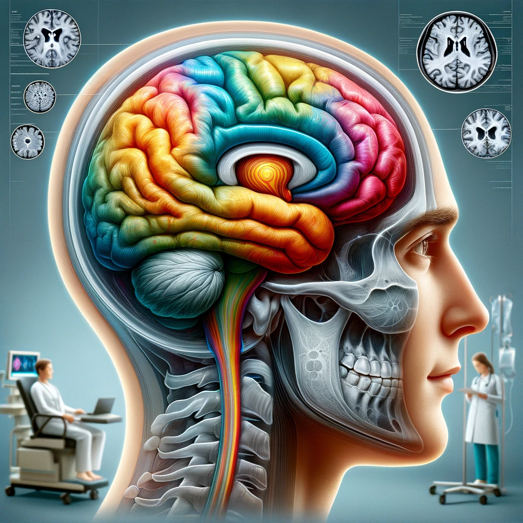
Radiologically, magnetic resonance imaging (MRI) plays a crucial role in the assessment of optic neuritis. In true cases of optic neuritis, MRI typically shows hyperintense lesions along the optic nerve on T2-weighted images and fat-suppressed sequences. Nevertheless, discrepancies may exist with false-positive cases, as imaging might display non-specific white matter lesions or changes that do not correspond to typical demyelination seen in actual optic neuritis.
An analysis of imaging findings in diagnosed false-positive cases reveals several noteworthy trends:
| Radiological Finding | Associated Condition | Frequency (%) |
|---|---|---|
| Non-specific White Matter Lesions | Migraine-associated changes | 30% |
| Optic Nerve Enhancement on MRI | Inflammatory diseases (e.g., sarcoidosis) | 20% |
| Infarcts or Vascular Changes | Transient Ischemic Attack (TIA) | 15% |
This table elucidates the importance of careful interpretation of MRI findings in those presenting with optic neuritis-like symptoms. The overlap of symptoms and imaging findings among various conditions can lead to diagnostic confusion. For instance, non-specific white matter lesions may arise from various causes, including vascular issues or chronic migraines—none of which are indicative of optic neuritis.
Furthermore, history-taking plays a pivotal role in distinguishing false-positive cases from actual optic neuritis. Detailed patient interviews that probe into the onset, duration, and progression of symptoms, along with prior medical history, can provide critical context that aids clinicians in making more informed decisions regarding diagnosis.
In sum, recognizing the clinical and radiological features associated with false-positive cases is critical in reducing misdiagnosis rates. By integrating patient histories, clinical presentations, and comprehensive imaging analysis, healthcare providers can significantly enhance diagnostic accuracy and improve patient outcomes in cases that initially seem to suggest optic neuritis.
Clinical and Radiological Features
Recommendations for Future Research
To enhance the accuracy of optic neuritis diagnoses and mitigate the prevalence of false-positive cases, future research should focus on several key areas. One major avenue is the exploration of advanced imaging techniques that can provide clearer delineation between true optic neuritis and other conditions that may masquerade as it. Techniques such as diffusion tensor imaging (DTI) and spectroscopy may yield additional insights into the pathology of the optic nerve and surrounding structures.
Moreover, longitudinal studies that track patients diagnosed with optic neuritis over time could prove invaluable. These studies should aim to correlate clinical symptoms with MRI findings over various stages of the disease, facilitating a deeper understanding of the characteristics that differentiate true optic neuritis from false positives. Such data could be instrumental in identifying specific biomarkers or clinical features that warrant differential diagnosis protocols.
Furthermore, it is essential to invest in clinician training programs that emphasize the nuances of optic neuritis diagnosis. By incorporating case studies on false-positive presentations into educational materials, healthcare professionals can be better equipped to recognize subtle variations in symptoms and imaging that could indicate alternative diagnoses.
Lastly, a collaborative approach that brings together neurologists, radiologists, and ophthalmologists could foster a more interdisciplinary understanding of optic neuritis. Multidisciplinary case reviews could promote the sharing of insights and experiences, leading to improved diagnostic strategies and management plans for patients presenting with optic neuritis-like symptoms.
A multifaceted research agenda that includes technological advancements, longitudinal studies, educational initiatives, and collaborative efforts is crucial for refining the understanding of optic neuritis and ultimately enhancing patient care.
Recommendations for Future Research
Future research initiatives must prioritize the establishment of standardized diagnostic criteria for optic neuritis that simultaneously account for the spectrum of conditions leading to false-positive diagnoses. Addressing this issue can facilitate a more accurate classification system, thus minimizing the potential for misdiagnosis.
In addition, the development of consensus guidelines among specialists in the field is essential. Variability in practice can contribute to inconsistencies in diagnosis; hence, a unified approach among neurologists, ophthalmologists, and radiologists regarding the interpretation of clinical findings and imaging results is necessary. Workshops and multidisciplinary meetings could serve as platforms to discuss novel insights, share clinical case studies, and establish best practices.
Moreover, there is a pressing need to investigate the genetic and environmental factors that may predispose certain individuals to develop conditions that mimic optic neuritis. Understanding the underlying etiology could lead to more individualized diagnostic strategies and potential preventative measures.
Another promising area for future exploration is the integration of artificial intelligence (AI) and machine learning technologies in imaging analysis. AI algorithms trained on vast datasets of MRI scans may enhance diagnostic precision by identifying patterns that might be overlooked by human observers. Implementing such technologies could streamline the diagnostic process and allow for timely and appropriate interventions for affected patients.
To underscore the significance of these initiatives, research metrics should be established to evaluate the impact of these strategies on diagnostic accuracy, patient outcomes, and healthcare resource utilization. Regular assessment and refinement of these interventions based on evolving evidence will be crucial in fostering a responsive and adaptive healthcare environment.
Ultimately, advancing our understanding of false-positive cases in optic neuritis through innovative research methods and collaborative frameworks will empower clinicians and improve the overall quality of care for patients presenting with this complex set of neurological symptoms.