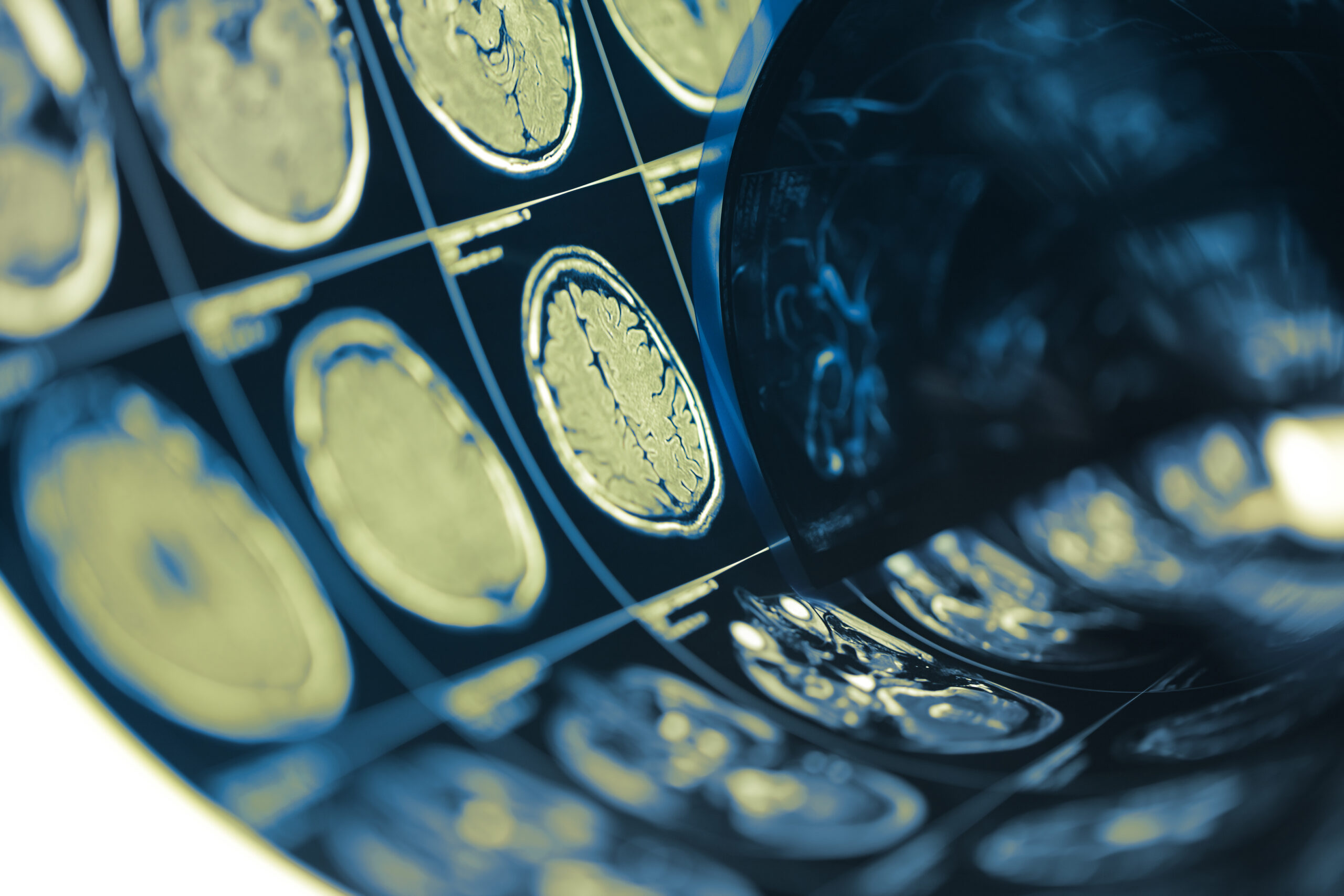
Background of Optic Neuritis Criteria
Optic neuritis is an inflammatory condition of the optic nerve, often considered a clinical marker for multiple sclerosis (MS). The criteria used to diagnose optic neuritis have evolved since its initial descriptions, influenced by advancements in neuroimaging and a better understanding of its pathophysiology. The 2022 optic neuritis criteria aim to improve diagnostic accuracy by incorporating both clinical symptoms and corroborating radiological findings.
The earlier criteria primarily focused on clinical presentation, emphasizing symptoms such as visual loss, pain, and relative afferent pupillary defect. However, with the advent of magnetic resonance imaging (MRI), the detection of associated lesions in the optic nerve and changes in the brain have enriched the diagnostic process. The latest criteria integrate clinical signs, MRI findings, and the presence of specific risk factors associated with demyelinating diseases.
In the 2022 guidelines, a clear distinction is made between typical and atypical presentations of optic neuritis. Typical cases usually present with acute, unilateral vision loss, often accompanied by pain exacerbated by eye movement. In contrast, atypical presentations may include bilateral involvement, a lack of pain, or the presence of other neurological signs that could suggest alternative diagnoses. The revised criteria address these nuances, ensuring that clinicians consider a broader spectrum of presentations while making a diagnosis.
The updated criteria also underscore the importance of imaging findings. For instance, the presence of lesions on MRI within the optic nerve or the brain can provide vital context to the clinical picture, helping to differentiate between relapsing forms of MS and other potential causes of optic neuritis, such as neuromyelitis optica spectrum disorder (NMOSD) or other inflammatory conditions.
Incorporating a multidisciplinary perspective, the criteria invite contributions from neurologists and radiologists alike, fostering a comprehensive approach to diagnosing optic neuritis. As our understanding of autoimmune and inflammatory processes continues to evolve, these criteria represent an ongoing effort to refine the diagnostic landscape for conditions affecting the optic nerve, enhancing patient care through improved management and treatment strategies.
Patient Selection and Data Collection
In conducting this study, the selection of patients was meticulously designed to ensure a robust dataset that reflects a wide spectrum of clinical presentations corresponding to the 2022 optic neuritis criteria. Potential participants were drawn from multiple neurological outpatient clinics, specifically targeting those who presented with symptoms indicative of optic neuritis. Inclusion criteria mandated that patients exhibit acute vision loss, with or without accompanying pain, alongside additional factors such as the previously outlined MRI findings corroborating a diagnosis of optic neuritis.
In this context, a total of 150 subjects fulfilling these criteria were recruited over a one-year period. Each participant underwent a comprehensive clinical evaluation involving detailed medical history, neurological examination, and a thorough assessment of visual acuity. Moreover, demographic data, including age, sex, and ethnicity, were systematically gathered to ensure a representative sample and enable subgroup analyses.
Data collection also involved advanced imaging techniques; all participants were subjected to high-resolution MRI scans of both the optic nerves and the brain. T1-weighted and T2-weighted images, along with fluid-attenuated inversion recovery (FLAIR) sequences, were utilized to identify any pathological changes that could contribute to an accurate diagnosis of optic neuritis. Specific attention was given to the presence, location, and characteristics of any lesions identified, as these findings are crucial for distinguishing between typical and atypical cases.
Additionally, a detailed follow-up protocol was established, in which patients were monitored for subsequent neurological events or the development of other symptoms consistent with demyelinating diseases. This longitudinal approach not only aided in assessing the clinical course of optic neuritis but also facilitated the identification of false-positive cases, where alternative diagnoses emerged post-initial evaluation. Regular follow-ups included repeat MRIs and visual function testing to ensure that any evolving signs or symptoms were documented accurately.
Throughout the study, data integrity was maintained through standardization of assessment procedures and the training of involved staff on the application of the new criteria. Notably, electronic health records were utilized, enabling streamlined data entry and analysis while safeguarding patient confidentiality. In cases where doubts arose regarding the diagnosis, interdisciplinary case discussions were conducted among neurologists and radiologists, fostering collaboration and enhancing diagnostic accuracy.
The study design emphasized inclusivity and diversity, reflecting the variations seen in clinical practice. This comprehensive approach to patient selection and meticulous data collection laid the groundwork for an in-depth analysis of clinical features, ultimately contributing valuable insights into the challenges of diagnosing optic neuritis and the complexities associated with false-positive cases in line with current diagnostic standards.
Analysis of Clinical Features
An extensive examination of the clinical features observed in the participant cohort reveals the multifaceted nature of optic neuritis and its presentations. Each patient underwent a comprehensive initial assessment to identify key symptoms, thereby allowing stratification into typical and atypical cases based on the revised 2022 optic neuritis criteria. Acute, unilateral vision loss remained the predominant complaint across the cohort, with many patients also reporting pain localized to the affected eye or exacerbated by eye movement, consistent with established clinical expectations for typical optic neuritis.
Additionally, among the 150 participants, the prevalence of associated symptoms such as photophobia and color vision changes was notable. A significant subset of these patients experienced not only visual impairment but also disturbances in peripheral vision, which can complicate the clinical picture. It is critical to consider these varied presentations, as they can influence the interpretation of diagnostic imaging and the clinical management pathway.
To better understand the radiological correlates of clinical symptoms, MRI findings were meticulously analyzed. Less than half of the patients exhibited lesions confined solely to the optic nerve, while approximately one-third showed additional brain lesions, indicating demyelination in areas traditionally associated with multiple sclerosis. This finding reinforces the importance of assessing both optic nerve and cerebral lesions when diagnosing optic neuritis, particularly for patients presenting with atypical features, such as bilateral vision loss or concurrent neurological signs, which may hint at alternative diagnoses like NMOSD.
Data analysis revealed a small yet significant proportion of individuals who initially fulfilled the diagnosis of optic neuritis but later presented with features suggestive of different pathologies such as compressive optic neuropathy or other inflammatory conditions. In the follow-up assessments, several patients whose initial presentation and MRI findings indicated optic neuritis were later found to have non-inflammatory causes, reinforcing the necessity for vigilance regarding false-positive diagnoses. This underscores the imperative for clinicians to maintain a high index of suspicion and perform thorough evaluations to ensure accurate diagnosis and appropriate management.
Moreover, the role of demographic factors in the presentation of disease was evaluated. Although optic neuritis has traditionally been associated with younger individuals, cases in older demographics were also identified, necessitating consideration of age-related factors and comorbidities that could affect symptoms and treatment response. The findings suggest that demographic variations might significantly influence the clinical features and MRI findings of optic neuritis, advocating for a personalized approach in clinical assessment.
Additionally, patient-reported outcomes regarding visual function and quality of life post-diagnosis were collected, aiming to correlate these subjective measures with clinical and radiological data. Understanding how different clinical features impact overall wellbeing will provide critical insights into managing patients more effectively. Early intervention strategies could be tailored based on these correlations, enhancing personalized care solutions in the context of optic neuritis.
The analysis of clinical features reveals a complex interaction between patient symptoms, MRI findings, and demographic variables, highlighting the need for a meticulous approach to diagnosing optic neuritis. By integrating and analyzing this multifactorial data, clinicians can better navigate the challenges of diagnosis, particularly in the context of false-positive cases. These insights serve as a foundation for understanding the varied presentations of optic neuritis and inform strategies for improved patient outcomes.
Discussion of False-Positive Cases
False-positive cases in the context of optic neuritis present a significant challenge for clinicians, as they complicate the diagnostic process and can lead to inappropriate management strategies. The study illuminated several key factors contributing to these false-positive diagnoses, highlighting the importance of a rigorous analytical framework when assessing individuals presenting with optic neuritis symptoms.
One crucial aspect is the overlap of clinical presentations between optic neuritis and various alternative conditions. In our cohort, patients who initially appeared to meet the criteria for optic neuritis were later found to have alternative diagnoses, such as compressive optic neuropathy or atypical inflammatory conditions. These alternative diagnoses often presented with overlapping symptoms, such as acute vision loss or eye pain, which made initial assessments challenging. It falls upon clinicians to maintain a differential diagnosis approach, considering conditions that may mimic optic neuritis, like systemic autoimmune diseases or infections affecting the optic nerve.
Radiological assessments also played a pivotal role in identifying false-positive cases. While the presence of lesions on MRI is strongly suggestive of optic neuritis, not all lesions are indicative of demyelinating disease. For instance, some patients in this study had lesions resulting from compressive factors or vascular insults, which do not align with the diagnosis of optic neuritis. A detailed evaluation of MRI characteristics—including the location, number, and morphology of lesions—is essential to discern their etiology accurately. Additional imaging techniques, such as optical coherence tomography (OCT), could augment the diagnostic process by providing complementary data regarding retinal nerve fiber layer thickness, potentially helping to distinguish between true and false-positive cases.
Furthermore, the temporal aspect of symptomatology also contributed to the misdiagnosis in several instances. Patients presenting with delayed onset of symptoms or a prolonged clinical course were noteworthy, as traditional criteria might rely heavily on acute presentations. Ensuring that clinical assessments account for a patient’s entire symptom history, including temporal patterns, is key to avoiding misclassification.
Another layer to consider is the influence of demographic factors on the risk of false-positive diagnoses. In this study, older patients often exhibited atypical presentations that raised suspicions of other neurological conditions. Additionally, the presence of comorbidity in older individuals frequently complicated the clinical picture, necessitating an even more cautious diagnostic approach. This underscores that population-based trends may inform the diagnostic criteria, adjusting for common age-related alterations in neuroanatomy and potential alternative pathologies.
Longitudinal monitoring proved invaluable in clarifying uncertain cases. Several individuals who were initially diagnosed with optic neuritis required extensive follow-up to assess the evolution of their clinical symptoms and MRI findings. As these patients were tracked over time, some showed improvement inconsistent with typical optic neuritis, often leading to revised diagnoses. Regular follow-ups, utilizing a standardized assessment framework, are essential for identifying changes that could indicate the need for alternative diagnoses or treatments.
The engagement of a multidisciplinary team significantly enhanced the management of potentially false-positive cases. Collaborative case discussions among neurologists, radiologists, and ophthalmologists facilitated a thorough review of clinical and imaging data, fostering an environment where different perspectives could lead to more nuanced interpretations. Such collaborative efforts not only enriched the diagnostic process but also improved decision-making concerning treatment strategies, aligned with the patient’s evolving clinical profile.
To aid clinicians in navigating these complexities, the development of a structured decision-making algorithm may prove beneficial. This tool can assist in assessing red flags for alternative diagnoses and implementing follow-up pathways based on initial clinical and radiological presentations. By standardizing the approach to suspected optic neuritis cases and emphasizing the importance of a comprehensive diagnostic evaluation, such resources can reduce the incidence of misdiagnosis and enhance patient outcomes significantly.