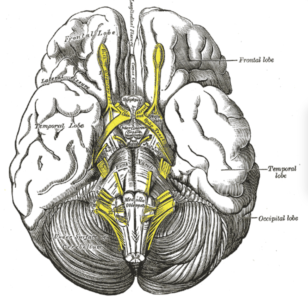
Functions of the Olfactory Nerve
The primary function of the olfactory nerve is to convey sensory information related to smell from the nasal cavity to the brain. This process begins when odorant molecules bind to receptors on the olfactory sensory neurons located in the olfactory epithelium. Each olfactory neuron expresses only one type of odorant receptor, and these receptors are tuned to detect specific chemicals. When an odorant binds to its receptor, it triggers a signal transduction pathway that results in the generation of an electrical signal, which travels along the axons of these neurons through the cribriform plate to the olfactory bulb[1].
In the olfactory bulb, these axons synapse with dendrites of second-order neurons, primarily mitral and tufted cells, within structures known as glomeruli. Each glomerulus receives input from olfactory sensory neurons that express the same receptor type, thus maintaining the specificity of odorant information. The mitral and tufted cells then relay this information deeper into the brain[1].
Olfactory Processes and Analysis into the Brain
Signal Transduction and Transmission
The initial phase of olfactory processing occurs at the level of the olfactory epithelium, where specific receptors bind odorants, leading to a cascade of cellular events that convert chemical signals into an electrical form. This electrical signal is transmitted via the olfactory nerve to the olfactory bulb[1].
Synaptic Integration in the Olfactory Bulb
Upon reaching the olfactory bulb, the signal is first processed in the glomerular layer, where the olfactory nerve fibers form synapses with the dendrites of mitral and tufted cells. This layer is characterized by heterogeneous expression of connexin 36, which forms gap junctions contributing to the synchronization of mitral and tufted cells, enhancing the reliability and speed of olfactory signal processing[1].
The olfactory bulb acts as a sophisticated processing unit that not only receives direct sensory input but also integrates modulatory feedback from higher brain centers. This integration helps in refining the sensory input based on past experiences, context, and the animal’s physiological state[4].
Higher Brain Processing
From the olfactory bulb, the processed information is transmitted to higher brain regions via the olfactory tract. Key areas involved include the piriform cortex, which is crucial for identifying and discriminating between different odors. The information then reaches the orbitofrontal cortex, where it is integrated with taste information to form flavor perception, and the amygdala, which is involved in emotional aspects of smell[2].
Additionally, olfactory signals are sent to the hippocampus and entorhinal cortex, areas important for memory and learning. This connection explains why certain smells can evoke vivid memories[2].
Neurotransmitter Systems
The olfactory system is also influenced by various neurotransmitter systems. For example, the serotonin 5-HT3 receptor is expressed in areas such as the hippocampus and amygdala, which interact with the olfactory system. The activation of these receptors can modulate the excitability of interneurons, thereby influencing olfactory processing and emotional responses to smells[2].
Conclusion
The olfactory nerve plays a critical role in detecting and processing smells, with its functions extending beyond mere odor detection to include significant roles in flavor perception, emotional responses, and memory recall. The intricate network of neurons and receptors, along with the complex synaptic integration in the olfactory bulb and the extensive projections to various brain regions, underscores the complexity and importance of olfactory processing in the brain. This system not only allows for the detection of environmental odors but also integrates these sensory inputs with cognitive and emotional centers, enriching the perceptual experience and guiding behavior.
Sources
[1] Heterogeneous expression of connexin 36 in the olfactory epithelium and glomerular layer of the olfactory bulb https://pubmed.ncbi.nlm.nih.gov/12687708/
[2] Nervous system distribution of the serotonin 5-HT3 receptor mRNA. https://pubmed.ncbi.nlm.nih.gov/8434003/
[3] Characterization of neuropeptide Y2 receptor protein expression in the mouse brain. I. Distribution in cell bodies and nerve terminals https://pubmed.ncbi.nlm.nih.gov/16998904/
[4] The neuron doctrine: a revision of functional concepts. https://www.ncbi.nlm.nih.gov/pmc/articles/PMC2591810/
[5] Illuminating the terminal nerve: Uncovering the link between GnRH-1 neuron and olfactory development. https://pubmed.ncbi.nlm.nih.gov/38488687/
[6] Evaluation of the Olfactory Nerve Transport Function by SPECT-MRI Fusion Image with Nasal Thallium-201 Administration https://pubmed.ncbi.nlm.nih.gov/21136183/
[7] [Clinical diagnosis of the olfactory nerve transport function]. https://pubmed.ncbi.nlm.nih.gov/23123717/
[8] Axon behavior in the olfactory nerve reflects the involvement of catenin–cadherin mediated adhesion https://pubmed.ncbi.nlm.nih.gov/17072833/pe around the olfactory bulb based on endoscopic cadaveric observations. https://pubmed.ncbi.nlm.nih.gov/38242165/
[19] Olfactory ensheathing cells and neuropathic pain https://www.ncbi.nlm.nih.gov/pmc/articles/PMC10201020/
[20] Visualization of the olfactory nerve using constructive interference in steady state magnetic resonance imaging https://pubmed.ncbi.nlm.nih.gov/27506829/

