Study Overview
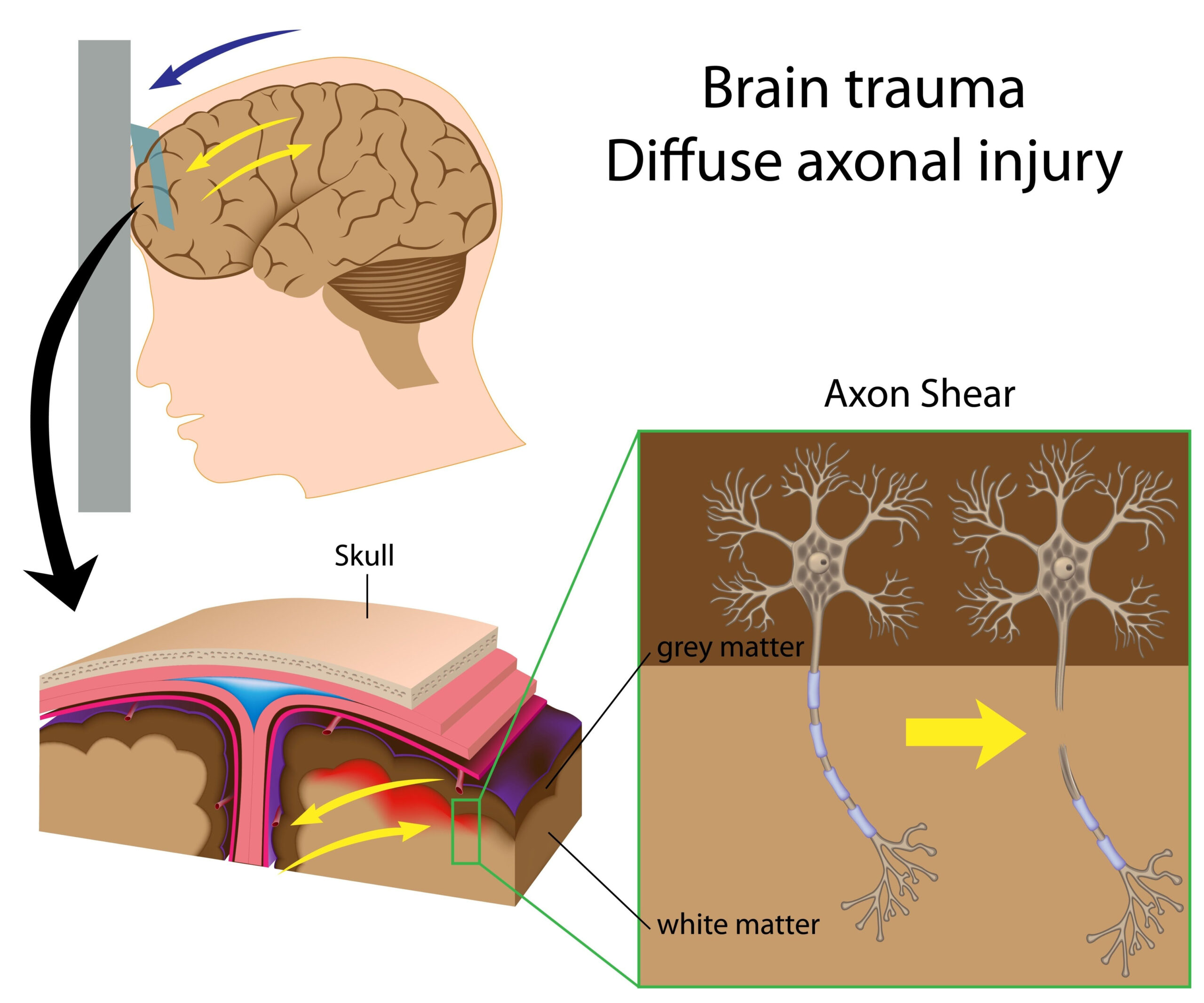
The research presented focuses on the development and validation of an advanced animal model aimed at simulating repetitive mild traumatic brain injury (rMTBI). This model is particularly significant for understanding the long-term consequences of such injuries, as they are prevalent in sports and military contexts. Traditional models often fell short in accurately replicating the complexities of human brain injuries, making it difficult for researchers to gain insights into the mechanisms of brain damage, recovery, and potential therapeutic interventions.
In this study, the authors implemented a modified approach that incorporates a thinned-skull window alongside fluid percussion injury techniques. This dual approach allows for the observation of the brain’s physiological response to injury under controlled conditions, closely mirroring the types of trauma experienced in real-life scenarios. By utilizing this innovative methodology, the researchers aimed to explore the cumulative effects of repeated mild injuries and the resultant neurobiological changes over time.
By creating a platform that not only simulates the initial impact but also permits ongoing assessment of brain function and structure, this study provides a crucial foundation for future investigations into treatment strategies for rMTBI. The ultimate goal is to enhance our understanding of the disorder, paving the way for improved clinical practices and interventions tailored to individuals at risk of recurrent head trauma.
Methodology
The study employed a carefully structured experimental approach to create an innovative animal model that accurately reflects the characteristics of repetitive mild traumatic brain injury (rMTBI). This model was developed to facilitate an in-depth examination of both the immediate and long-term effects of mild brain injuries, ultimately aiming to yield insights that traditional models have failed to deliver.
Initially, the researchers selected a suitable animal subject, specifically adult mice, chosen for their robust response to traumatic brain injury and their well-documented neurobiological characteristics. Prior to any experimental manipulations, the animals underwent a series of pre-surgical evaluations to establish baseline neurobehavioral function and ensure their overall health.
In a critical innovation, the study utilized a thinned-skull window technique. This procedure involved carefully reducing the thickness of the skull over specific cortical regions without breaching the dura mater, the protective layer surrounding the brain. This method not only allows for better visualization of the brain during subsequent injury of the fluid percussion type but also minimizes additional trauma that could confound results. The thinned-skull window facilitates ongoing monitoring of brain activity and structural changes through advanced imaging techniques.
For the fluid percussion injury (FPI) component, the researchers implemented a precise protocol whereby a controlled fluid wave was applied to the exposed brain through the thinned skull. This approach mimics the forces experienced during incidental impacts, common in both athletic and military settings. The timing, pressure, and frequency of the pulses were meticulously calibrated to produce mild injuries, thereby avoiding severe damage while still invoking the physiological responses characteristic of rMTBI.
Repeated injury regimens were established, with intervals between impacts allowing for observation of potential accumulated damage or recovery indicators. Following the induction of injury, the animals were monitored for behavioral changes using established neurobehavioral assessments, which included evaluations of motor function, memory retention, and anxiety-like behaviors.
Post-injury analysis involved employing histological techniques to assess brain tissue response. This included staining methods for detecting neuroinflammation, cellular apoptosis, and neurogenesis. Additionally, advanced imaging modalities, such as MRI and CT scans, were utilized to visualize changes in brain morphology over time, delivering quantitative data that could correlate with the behavioral assessments.
In summary, this meticulous and multi-faceted methodology not only provided a groundbreaking framework to study rMTBI but also opened avenues for real-time observation of brain function and morphology. The integration of innovative surgical techniques, advanced imaging, and rigorous behavioral assessments creates a comprehensive analytical approach to better understand the dynamic nature of repetitive mild brain injuries and their broader implications.
Key Findings
The findings from the study reveal several significant insights into the physiological and behavioral consequences of repetitive mild traumatic brain injuries (rMTBI) as modeled in the modified mouse model. Key outcomes of the research are centered on neurobehavioral changes, neuroinflammatory responses, and structural alterations in the brain, providing a multidimensional understanding of rMTBI.
Firstly, the behavioral assessments demonstrated marked changes following the induction of rMTBI. Mice subjected to repeated mild injuries exhibited notable impairments in motor coordination, memory performance, and increased anxiety-like behaviors compared to the control group. These findings underscore the potential for cumulative damage resulting from mild traumatic brain injuries, even in the absence of overt severe trauma. The observed motor deficits were quantified through tasks such as balance beam evaluations and rotarod tests, indicating a clear degradation in motor skills which may mirror challenges faced by human athletes and soldiers post-trauma.
In addition to behavioral manifestations, histological analyses revealed critical neurobiological changes within the brain tissue. High levels of neuroinflammation were detected in the affected cortical regions, evidenced by an increase in pro-inflammatory cytokines and activated microglia. This suggests that even mild injuries can instigate significant inflammatory cascades, thus playing a vital role in the pathology associated with rMTBI.
Moreover, the study revealed alterations in neurogenesis and cellular apoptosis within the hippocampus and other key brain areas responsible for cognitive function. The presence of apoptotic cells indicated that rMTBI led to neuronal loss, potentially contributing to the observed deficits in learning and memory. The reduced neurogenic capacity, as shown by diminished markers of new neuron formation, highlights a long-term alteration in brain plasticity, suggesting sustained effects of mild injuries that extend beyond the acute phase.
Imaging results further enriched our understanding of structural changes resulting from rMTBI. MRI scans indicated that the integrity of the blood-brain barrier was compromised, with increased permeability observed in the post-injury phase. This disruption raises concerns about the potential for secondary injury responses and the role of neurovascular dysfunction in chronic outcomes associated with repeated head trauma.
Overall, these findings contribute to a more nuanced model that reflects the complexities associated with rMTBI. The integrative approach, combining behavioral, histological, and imaging modalities, provides a robust framework for understanding the multifaceted nature of mild brain injuries. This work could pave the way for future studies aimed at identifying therapeutic interventions and preventative strategies for individuals who risk repeated head injuries, particularly in vulnerable populations such as athletes and military personnel.
Clinical Implications
The implications of this study for clinical practice are profound, as they deepen our understanding of the repercussions associated with repetitive mild traumatic brain injuries (rMTBI). As awareness surrounding the long-term consequences of head injuries grows, this research underscores the necessity for heightened surveillance and proactive management strategies for individuals at risk, particularly among athletes and military personnel.
Firstly, the behavioral changes observed in the modified mouse model—including impairments in motor coordination, memory, and increased anxiety—parallel symptoms commonly reported in humans post-concussion. These findings may prompt clinicians to reassess current concussion protocols, emphasizing the need for rigorous follow-ups and cognitive evaluations even after mild head injuries. The absence of severe trauma does not preclude significant neurobehavioral impacts, suggesting that incremental and cumulative injuries should not be dismissed simply because initial assessments yield no overt signs of damage.
Furthermore, the presence of neuroinflammatory markers indicated a biological response that could have significant implications for therapeutic approaches. Inflammation within the brain, triggered by repeated mild trauma, may play a critical role in augmenting neurodegenerative processes over time. This discovery points to the potential utility of anti-inflammatory interventions in managing patients who have sustained rMTBI. Such treatments could mitigate the inflammatory cascades initiated by head injuries, potentially preserving cognitive function and reducing the risk of long-term neurodegeneration.
The study also highlights alterations in neurogenesis and increased instances of neuronal apoptosis. This raises vital questions concerning rehabilitation strategies, as fostering neuroplasticity and promoting neurogenesis could be critical components in recovery. Current therapy regimens may need to be expanded to incorporate neuroprotective agents and cognitive training exercises designed to bolster brain plasticity, enhancing recovery processes after injury.
Additionally, the observed disruption of the blood-brain barrier (BBB) calls for an urgent reevaluation of how brain injuries are diagnosed and treated. Clinically, understanding how rMTBI can compromise the integrity of the BBB highlights the importance of monitoring and managing secondary injury risks. This may involve employing imaging techniques to assess BBB integrity in individuals with a history of head trauma, offering a biomarker for ongoing risk assessment and intervention strategies.
Given the findings concerning cumulative damage, personalized management approaches that take into account an individual’s history of head trauma could be beneficial. Sports organizations and military practice could implement tailored protocols that involve monitoring for neurobehavioral changes over extended periods, seeking to identify at-risk individuals early and providing them with appropriate support.
In summary, the insights gleaned from this animal model are not merely academic; they present tangible pathways for enhancing the clinical handling of rMTBI. By translating these findings into practice, healthcare providers can better inform athletes and military personnel about the risks of repeated head trauma and develop interventions that clarify the long-term implications of even mild injuries. This concerted effort could lead to more effective preventive measures and treatment strategies, greatly benefitting those susceptible to the effects of recurrent mild traumatic brain injuries.


