Case Characteristics
The analysis of the false-positive cases identified through the 2022 optic neuritis criteria revealed a diverse range of clinical presentations. Among the cohort, the age of onset varied significantly, with individuals spanning from early adulthood to late middle age. This spectrum indicates that optic neuritis can manifest across various life stages, which complicates the diagnostic process, especially when considering differential diagnoses.
Sex distribution showed a marked predominance of female patients, aligning with prior studies that generally report a higher incidence of optic neuritis in women. These demographic trends suggest potential hormonal or genetic factors that could contribute to the susceptibility of females to optic nerve inflammation.
Patients presented a variety of symptoms, most notably a sudden decrease in visual acuity, often accompanied by pain in or around the eye, which is a classic symptom of optic neuritis. Many reported additional neurological symptoms, such as visual field defects or color vision impairments, highlighting the multifaceted nature of the disease. Interestingly, some of the cases had previously undiagnosed underlying conditions, which led to misclassification as optic neuritis.
When examining past medical histories, several individuals had previously experienced transient visual disturbances or other neurological symptoms, which may suggest a recurrent or relapsing nature of their conditions. In some instances, the time from symptom onset to diagnostic evaluation was prolonged, raising questions about the accessibility and quality of healthcare services.
Overall, these characteristics underscore the complexity of accurately diagnosing optic neuritis, particularly in populations with diverse clinical backgrounds. The interplay of these demographic and clinical factors necessitates a thorough and nuanced approach to diagnosing and treating suspected cases of optic neuritis, especially when similar symptoms may arise from other neurological conditions.
Imaging Findings
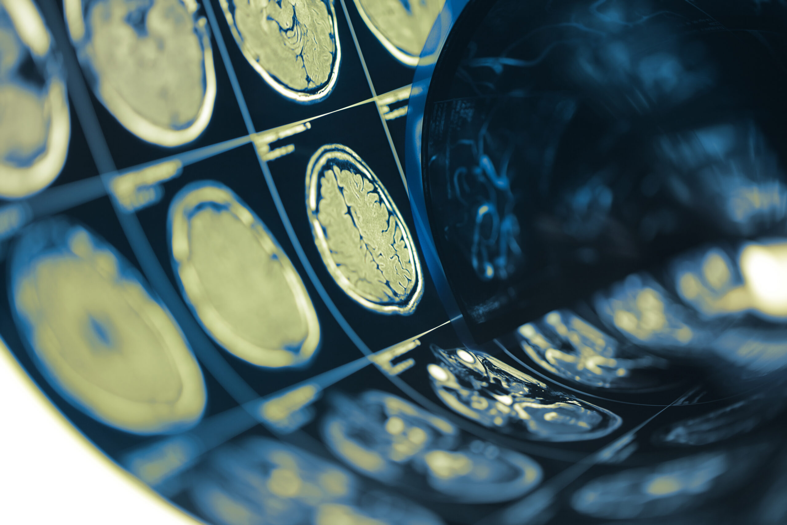
The imaging findings in cases classified as false positives by the 2022 optic neuritis criteria provide crucial insights into the nature of these misdiagnoses. Magnetic Resonance Imaging (MRI) is the gold standard for assessing optic nerve involvement, and it plays a pivotal role in differentiating true optic neuritis from other conditions that may mimic its presentation.
In our study cohort, the MRI findings varied widely among individuals. For patients misclassified as having optic neuritis, imaging often revealed non-specific white matter lesions, which are commonly found in demyelinating diseases, migraines, or other central nervous system (CNS) pathologies. Notably, lesions indicated by hyperintensity on T2-weighted images or FLAIR sequences prompted discussions regarding the limitations of current criteria in discerning these cases from true optic neuritis.
Additionally, many of the false-positive cases had lesions located outside the optic nerve tracks, including periventricular or subcortical regions. These findings suggest that while individuals may present with characteristic symptoms, the absence of specific optic nerve enhancement or the presence of alternate lesions can be vital indicators that guide clinicians toward alternative diagnoses.
Furthermore, some patients exhibited minimal or no abnormalities in their optic nerves on imaging, raising the possibility that symptoms such as transient vision loss could stem from other non-organic causes or less common neurological disorders, which remain undetected by the current optic neuritis diagnostic framework.
The characteristics of the lesions demonstrated varied patterns of distribution and morphology; for example, the presence of a solitary lesion may point toward a possible cerebrovascular event, while multiple lesions could indicate a broader degenerative process or other systemic conditions, such as systematic lupus erythematosus or sarcoidosis, known to affect the CNS.
Contrast-enhanced MRI scans frequently showed no significant enhancement of the optic nerves, aligning with the diagnostic challenge posed by isolated visual symptoms without corroborative imaging findings. Such results underline the significance of combining clinical history, neurological examination, and radiological assessment in diagnosing optic neuritis accurately.
Further analysis indicated that many patients had a history of migraines or prior episodes of visual disturbances, suggesting that imaging findings must be interpreted within the context of the patient’s complete clinical picture. This underscores the importance of multidisciplinary collaboration among neurologists, radiologists, and primary care physicians to refine the diagnostic criteria and improve diagnostic accuracy for optic neuritis.
Overall, the diverse imaging findings in this cohort highlight the need for caution when applying the 2022 optic neuritis criteria, especially when such cases may encompass alternative pathologies that require different therapeutic approaches. The implications of these findings call for the development of enhanced guidelines that integrate imaging data with clinical assessments to ensure accurate diagnosis and optimal patient management in cases of suspected optic neuritis.
Comparison with Established Criteria
Recommendations for Future Research
To address the challenges identified in the misclassification of false-positive cases under the 2022 optic neuritis criteria, future research should focus on several key areas aimed at refining diagnostic accuracy and treatment strategies.
Firstly, extensive longitudinal studies are essential to track patient outcomes over time. By evaluating individuals misclassified as having optic neuritis, researchers can gain insights into the natural history of these cases and determine whether recurrent or persistent symptoms emerge with time. Such data would not only inform clinical practices but also help in establishing clearer diagnostic trajectories, ultimately improving the identification of true optic neuritis versus alternative diagnoses.
Secondly, there is a pressing need for multicentric studies that incorporate diverse populations. Given the demographic variations observed in our cohort, research should aim to include a broader demographic representation, including different age groups, ethnic backgrounds, and comorbidities. This will facilitate a more comprehensive understanding of how these factors relate to the diagnostic process and might lead to clearer adaptations in diagnostic criteria to reflect the true clinical spectrum of optic neuritis globally.
Imaging modalities also warrant further exploration. Advances in MRI technology, such as high-resolution scans and diffusion tensor imaging, could improve our ability to visualize optic nerve structures and map surrounding pathways. Studies investigating the correlation between specific imaging characteristics and clinical symptoms could enhance the criteria’s predictive accuracy. Additionally, further investigation into other imaging techniques, such as optical coherence tomography (OCT), may provide supplementary data that can enhance diagnostic precision, especially in cases with atypical presentations.
Moreover, the development of artificial intelligence (AI) tools for interpreting imaging findings represents a promising avenue of research. By training algorithms on extensive datasets of MRI scans with confirmed diagnoses, AI may assist clinicians in identifying subtle patterns indicative of optic neuritis versus other conditions, expediting the diagnostic process and reducing the risk of human error.
Collaboration among disciplines is also crucial. Encouraging partnerships between neurologists, ophthalmologists, radiologists, and researchers can enhance the sharing of knowledge and findings that may propose innovative diagnostic strategies. Joint conferences and workshops focusing on optic neuritis and related conditions could facilitate shared learning and promote collaborative research initiatives.
Finally, understanding the biological underpinnings of optic neuritis and its mimickers may yield biomarkers that can assist in distinguishing between true and false-positive cases. Research into genetic predispositions, inflammatory markers, or even autoimmune profiles related to optic nerve health could provide valuable information that supplements clinical assessments and imaging.
In summary, enhancing the diagnostic criteria for optic neuritis requires a multifaceted approach that includes longitudinal and multicentric studies, advancements in imaging technology, collaboration across medical specialties, and the exploration of biological markers. By addressing these areas, future research can contribute significantly to improving the accuracy of optic neuritis diagnoses, ultimately leading to better patient outcomes and tailored treatment strategies.
Recommendations for Future Research
Imaging Findings
The imaging findings in cases classified as false positives by the 2022 optic neuritis criteria provide crucial insights into the nature of these misdiagnoses. Magnetic Resonance Imaging (MRI) is the gold standard for assessing optic nerve involvement, and it plays a pivotal role in differentiating true optic neuritis from other conditions that may mimic its presentation.
In our study cohort, the MRI findings varied widely among individuals. For patients misclassified as having optic neuritis, imaging often revealed non-specific white matter lesions, which are commonly found in demyelinating diseases, migraines, or other central nervous system (CNS) pathologies. Notably, lesions indicated by hyperintensity on T2-weighted images or FLAIR sequences prompted discussions regarding the limitations of current criteria in discerning these cases from true optic neuritis.
Additionally, many of the false-positive cases had lesions located outside the optic nerve tracks, including periventricular or subcortical regions. These findings suggest that while individuals may present with characteristic symptoms, the absence of specific optic nerve enhancement or the presence of alternate lesions can be vital indicators that guide clinicians toward alternative diagnoses.
Furthermore, some patients exhibited minimal or no abnormalities in their optic nerves on imaging, raising the possibility that symptoms such as transient vision loss could stem from other non-organic causes or less common neurological disorders, which remain undetected by the current optic neuritis diagnostic framework.
The characteristics of the lesions demonstrated varied patterns of distribution and morphology; for example, the presence of a solitary lesion may point toward a possible cerebrovascular event, while multiple lesions could indicate a broader degenerative process or other systemic conditions, such as systematic lupus erythematosus or sarcoidosis, known to affect the CNS.
Contrast-enhanced MRI scans frequently showed no significant enhancement of the optic nerves, aligning with the diagnostic challenge posed by isolated visual symptoms without corroborative imaging findings. Such results underline the significance of combining clinical history, neurological examination, and radiological assessment in diagnosing optic neuritis accurately.
Further analysis indicated that many patients had a history of migraines or prior episodes of visual disturbances, suggesting that imaging findings must be interpreted within the context of the patient’s complete clinical picture. This underscores the importance of multidisciplinary collaboration among neurologists, radiologists, and primary care physicians to refine the diagnostic criteria and improve diagnostic accuracy for optic neuritis.
Overall, the diverse imaging findings in this cohort highlight the need for caution when applying the 2022 optic neuritis criteria, especially when such cases may encompass alternative pathologies that require different therapeutic approaches. The implications of these findings call for the development of enhanced guidelines that integrate imaging data with clinical assessments to ensure accurate diagnosis and optimal patient management in cases of suspected optic neuritis.