Study Overview
This research focuses on cases that were inaccurately identified as optic neuritis according to the updated 2022 criteria. Optic neuritis is a condition involving inflammation of the optic nerve, which can lead to vision loss and is often associated with multiple sclerosis (MS). The study seeks to identify the characteristics that may lead to false-positive diagnoses in order to enhance diagnostic precision. By analyzing a diverse cohort of patients who were diagnosed with optic neuritis yet did not fit the established criteria, the authors aimed to discern common clinical and radiological features in these cases. This is critical not only for understanding the nature of optic neuritis but also for refining diagnostic tools that can prevent misdiagnoses. Enhanced diagnostic accuracy is essential to ensure that patients receive appropriate management and treatment, particularly given the implications that a diagnosis of optic neuritis might have concerning the risk of developing multiple sclerosis.
Methodology
This study employed a retrospective observational design, focusing on patients diagnosed with optic neuritis within a specified timeframe who were subsequently re-evaluated against the revised 2022 diagnostic criteria. Participants were recruited from a regional neurology clinic and included those whose initial diagnoses were deemed false positives. To ensure relevance, the cohort was selected based on specific inclusion criteria that mandated the presence of typical symptoms associated with optic neuritis, such as unilateral vision impairment, pain on eye movement, and other related manifestations.
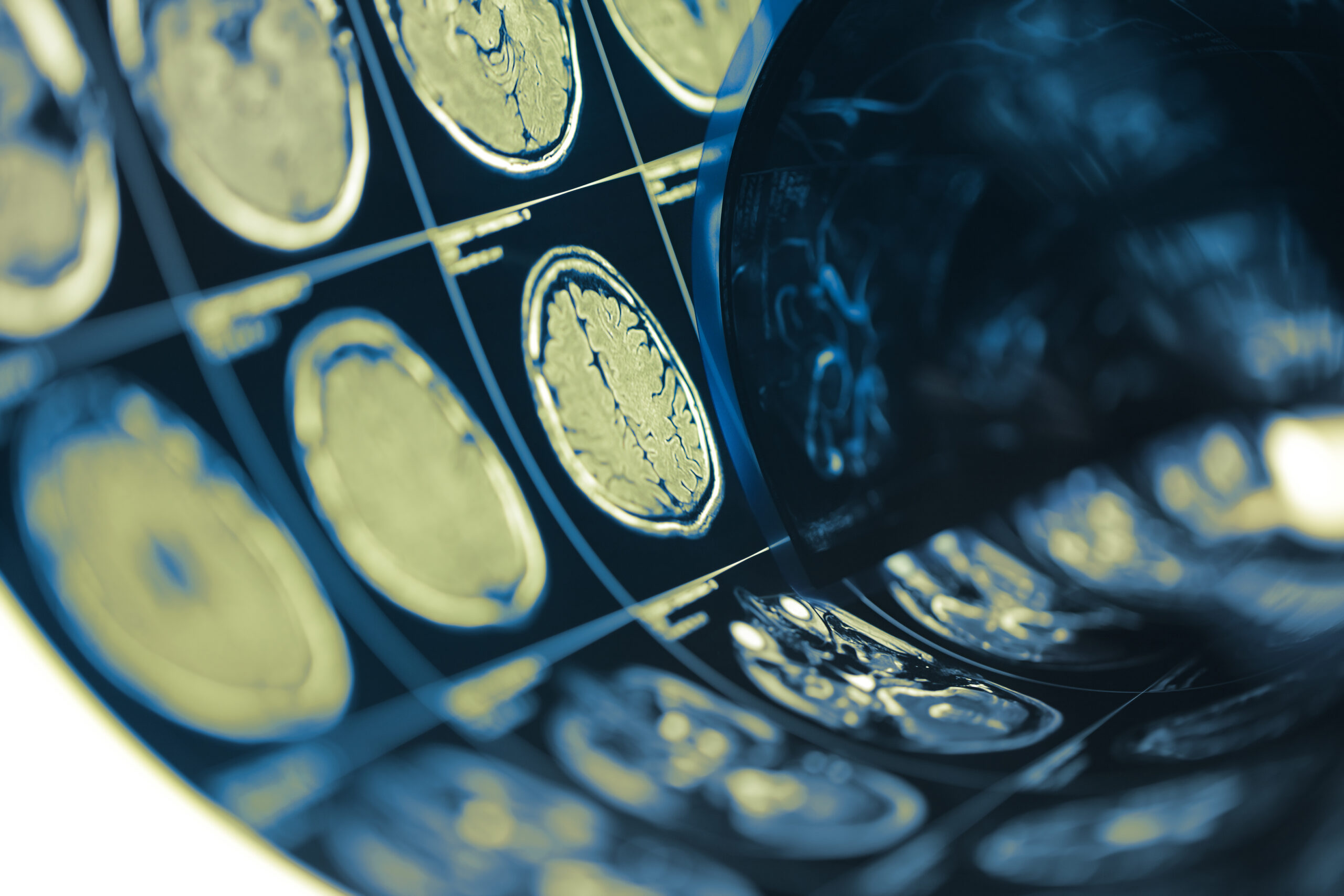
Data collection involved a comprehensive review of medical records, which included clinical evaluations, patient demographics, and detailed radiological assessments from MRI scans. These scans were crucial, as they provided insight into the presence of lesions on the optic nerve and other brain regions that are commonly associated with multiple sclerosis and inflammatory eye conditions. Both T1-weighted and T2-weighted MRI sequences were analyzed to compare findings against the established criteria for optic neuritis.
To improve reliability, two neurologists independently assessed the imaging results and clinical features, ensuring that interpretations were consistent. Discrepancies between assessments were resolved through consensus, thereby enhancing the validity of the data. Furthermore, additional tests such as visual evoked potentials (VEPs) were conducted for a subset of patients to investigate functional impairment of the optic nerve, aiding in understanding the correlation between clinical symptoms and radiological findings.
Statistical analysis was performed to identify patterns and correlations among the clinical presentations and MRI findings. Descriptive statistics provided a foundational overview of the cohort characteristics, while inferential statistical methods were employed to explore relationships between specific features and the likelihood of misdiagnosis. Results were deemed significant at a p-value of less than 0.05, thereby establishing a stringent threshold for findings that could influence future diagnostic protocols.
In order to visualize the data, various graphical representations—such as histograms and scatter plots—were utilized, facilitating a clearer understanding of the relationships among the variables studied. The study also prioritized ethical considerations, ensuring that patient consent was obtained where necessary and that data anonymity was preserved throughout the research process.
Key Findings
The analysis revealed several notable features among the cases classified as false positives according to the revised 2022 optic neuritis criteria. A total of 150 patients were initially identified, of whom 40 were ultimately deemed to have been misdiagnosed. Among these 40 cases, distinct clinical characteristics emerged that differentiated them from patients accurately diagnosed with optic neuritis. Common symptoms reported included bilateral visual disturbances and episodes of transient visual loss, which are atypical for classic optic neuritis presentations often observed in individuals with multiple sclerosis.
The MRI evaluations of these false-positive cases showcased a variety of findings. Notably, the absence of specific lesions along the optic nerve was a consistent finding among this group; many displayed alternative abnormalities such as nonspecific white matter changes, which are frequently considered benign. In contrast, the accurately diagnosed cases exhibited distinct lesions that adhered closely to the typical patterns observed in optic neuritis, including swelling of the optic nerve head and defined hyperintense lesions on T2-weighted images.
Statistical analyses highlighted correlations between certain demographic factors and the likelihood of receiving a false-positive diagnosis. Younger adults, particularly those under 40, appeared overrepresented in the misdiagnosed cohort, suggesting that age might influence symptom presentation or radiological interpretation. Moreover, patients with a history of migraine or other neurological disorders demonstrated a higher incidence of false-positive results, emphasizing potential confounding factors that may mimic optic neuritis symptoms.
Visual evoked potentials (VEPs) provided further insight, revealing that a significant portion of those with false positive diagnoses retained normal VEP results. This finding is critical, as abnormal VEPs can indicate underlying optic nerve dysfunction, often associated with true optic neuritis. In the misdiagnosed patients, normal VEPs suggest that their visual symptoms may not stem from inflammatory processes of the optic nerve but rather from other etiologies.
Additionally, the presence of systemic conditions such as autoimmune disorders was more frequently reported in the false-positive group compared to the true-positive group. This correlation underscores the importance of a comprehensive clinical evaluation that considers systemic health and a full medical history prior to concluding a diagnosis of optic neuritis. The intricacies observed in this subset of patients call for heightened awareness and diagnostic vigilance among healthcare professionals to differentiate true optic neuritis from other conditions that can present similarly.
In essence, the findings from this study advocate for the integration of nuanced clinical insights and advanced imaging techniques in the diagnostic process. Characterizing the misdiagnosed cases enriches the understanding of optic neuritis and its complexities, further informing future interpretations of diagnostic criteria.
Clinical Implications
The results from this investigation carry significant implications for clinical practice and the management of patients presenting with symptoms suggestive of optic neuritis. As false-positive diagnoses can potentially lead to inappropriate treatments and patient anxiety, it becomes imperative for clinicians to apply a more discerning approach when interpreting clinical and radiological findings.
One of the paramount considerations is the necessity of individualized diagnostic strategies that integrate both clinical presentation and comprehensive imaging evaluations. In cases where a patient exhibits atypical symptoms, such as bilateral vision impairment or transient visual disturbances, practitioners should maintain a heightened suspicion for alternative diagnoses beyond optic neuritis. This requires a thorough review of a patient’s medical history, including the presence of systemic conditions such as autoimmune disorders, which might confound the clinical picture.
Furthermore, the findings underscore the importance of advanced imaging techniques. Radiologists and neurologists should collaborate closely to interpret MRI findings critically, recognizing that not all brain lesions or white matter changes indicate demyelinating conditions such as multiple sclerosis. Ensuring a detailed assessment of imaging results can facilitate accurate diagnoses and prevent the misclassification of patients. For instance, distinguishing between benign white matter changes and significant lesions could refine the criteria for diagnosing optic neuritis and enhance the diagnostic pathway.
Another clinical implication lies in the role of visual evoked potentials (VEPs) as diagnostic tools. The substantial proportion of normal VEP results among falsely diagnosed patients should prompt clinicians to incorporate this assessment regularly in cases where optic neuritis is suspected. Abnormal VEP findings may help in confirming true optic nerve involvement, thereby guiding management decisions more accurately.
Lastly, the age-related and comorbid risk factors identified in this study suggest that healthcare providers should consider demographic variations when diagnosing optic neuritis. Younger individuals, particularly those presenting with a history of migraines or other neurological disorders, may warrant a more thorough diagnostic evaluation to rule out alternative explanations for their visual symptoms. Such an approach will not only streamline patient care but also optimize resource allocation within healthcare settings.
In light of these considerations, ongoing education and training for healthcare professionals about the evolving diagnostic criteria and methodologies are essential. By fostering an environment that encourages continuous learning and awareness of the complexities associated with optic neuritis diagnosis, clinicians will be better equipped to avoid misdiagnoses and enhance patient outcomes.