Mechanical Mechanisms of Brain Damage
Brain damage in conditions like idiopathic normal pressure hydrocephalus (iNPH) has been increasingly understood through the lens of biomechanics. iNPH manifests primarily in older adults and is characterized by specific symptoms such as gait disturbances, cognitive decline, and urinary incontinence. The underlying mechanical mechanisms that lead to such damage are often linked to the abnormal accumulation of cerebrospinal fluid (CSF) in the brain’s ventricles, which can lead to increased intracranial pressure and altered brain morphology.
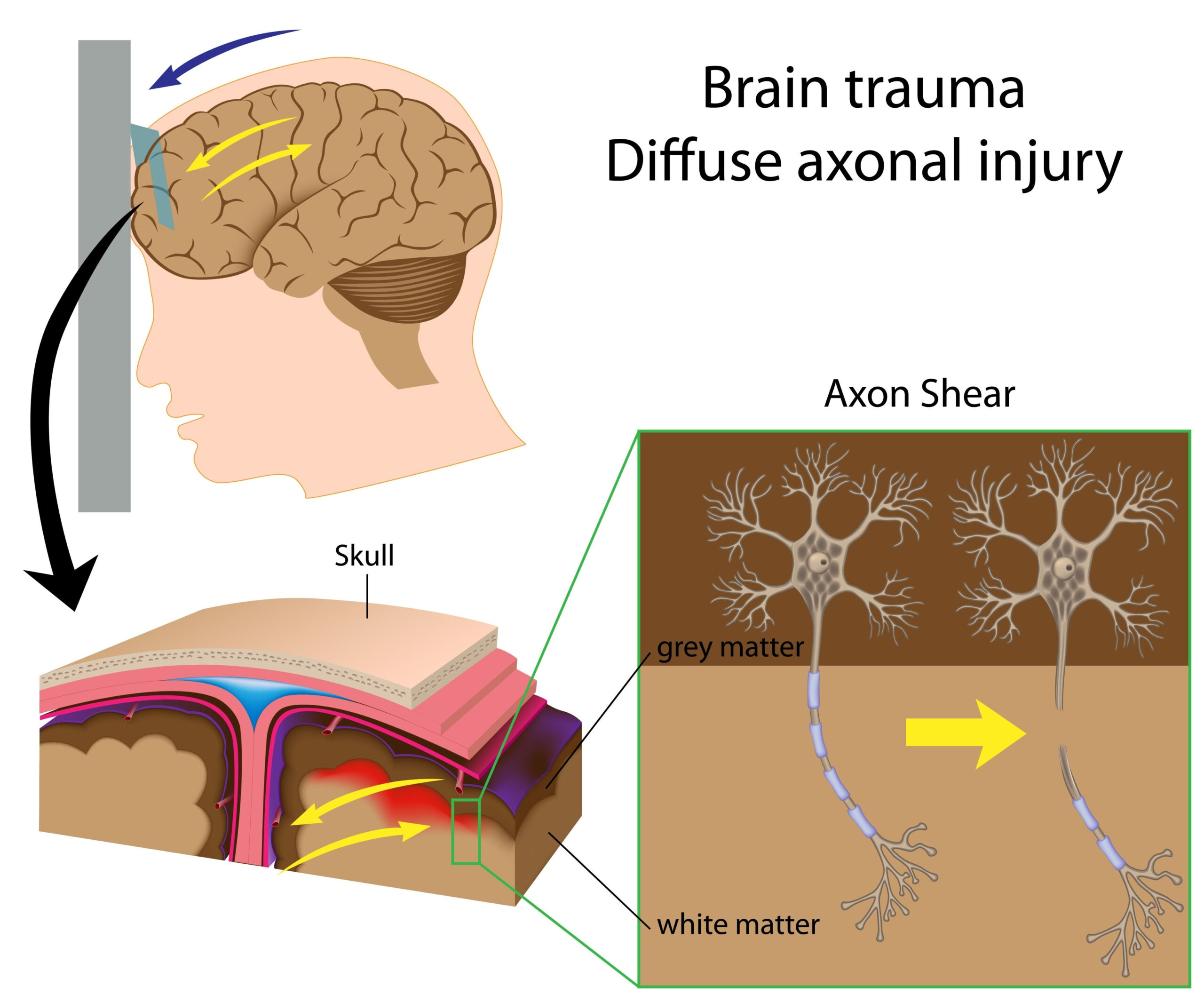
As CSF accumulates, the resulting ventricular enlargement exerts pressure on surrounding brain tissue, impacting its normal structure and function. This pressure can disrupt the delicate balance of cellular processes in the brain, leading to neuronal damage and loss. Studies have shown that the mechanical stress resulting from this pressure can induce cellular responses that contribute to neurodegeneration over time. Specifically, neurons may experience changes in their morphology due to deformation caused by the mechanical forces exerted by the enlarged ventricles. These changes can impact overall neuronal health, potentially leading to cell death and the associated cognitive and motor deficits observed in patients.
Moreover, the mechanical environment of the brain is not only influenced by CSF dynamics but also by the properties of brain tissue itself. The stiffness of brain tissue, which can vary due to aging and disease, plays a significant role in how brains respond to the changing forces. When brain tissue is subjected to increased pressure or mechanical forces, it can lead to further complications, such as vascular damage or inflammation. Research suggests that this interplay between CSF dynamics and the biomechanical properties of brain tissue is critical in understanding how brain damage occurs.
Advancements in imaging technologies and biomechanical modeling have enabled researchers to simulate and visualize these mechanical interactions more effectively. By applying patient-specific data to these models, it becomes possible to examine how variations in individual anatomy can influence the susceptibility to brain damage in conditions like iNPH. This approach allows for a more nuanced understanding of the disease process, potentially leading to better-targeted interventions for patients.
In summary, the mechanical mechanisms that contribute to brain damage in idiopathic normal pressure hydrocephalus involve a complex interplay between enlarged ventricular size, altered pressure dynamics, and the biomechanical properties of brain tissue. Understanding these mechanisms is essential for developing effective therapeutic strategies and improving patient outcomes.
Patient-Informed Approach
In addressing the complexities of idiopathic normal pressure hydrocephalus (iNPH), a patient-informed approach has emerged as a pivotal strategy for enhancing both the understanding and management of this condition. This methodology centers on incorporating patient-specific data into research and clinical practice, thereby allowing for a more personalized examination of the biomechanical interactions at play in each individual case.
At the heart of the patient-informed approach is the recognition that the variability among patients is significant. Factors such as age, gender, health status, and individual anatomical differences can all influence how the brain responds to the accumulation of cerebrospinal fluid (CSF) and the resulting mechanical stress on brain tissue. For instance, some patients may exhibit pronounced symptoms despite similar ventricular enlargement as those who have mild or no symptoms. This discrepancy underscores the need for tailored assessments that consider the unique characteristics of each patient’s condition.
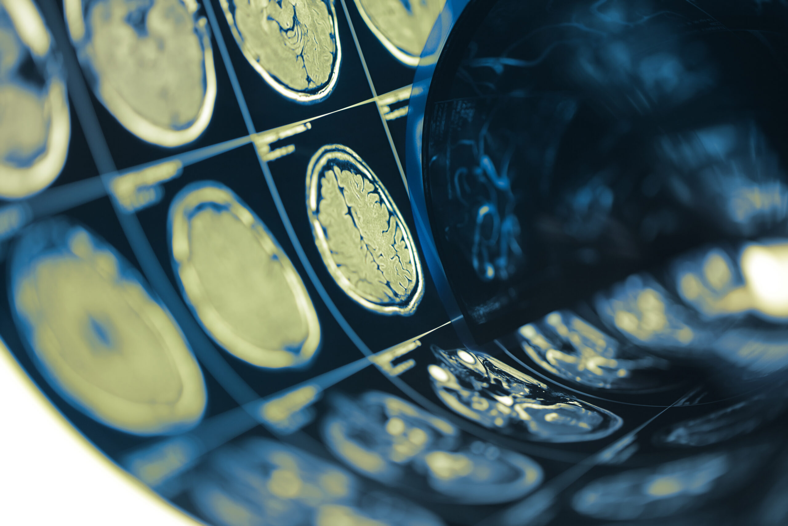
To implement this approach effectively, comprehensive data collection is essential. Researchers can utilize advanced imaging techniques, such as magnetic resonance imaging (MRI), to capture detailed snapshots of individual brain morphology and CSF dynamics. These images provide vital information that can be integrated into biomechanical models to simulate patient-specific scenarios. For example, analyses can reveal how variations in ventricular size and brain tissue stiffness impact mechanical stress distribution across different regions of the brain.
Engaging patients in the research process not only enhances data quality but also empowers them as active participants in their own care. Surveys and questionnaires can be employed to gather patient-reported outcomes that reflect how they experience their symptoms, which may include cognitive decline and gait disturbances. This subjective data is crucial, as it helps to correlate clinical findings with the actual lived experiences of patients, leading to a more holistic understanding of the condition.
Moreover, collaboration with multidisciplinary teams involving neurologists, radiologists, biomechanical engineers, and, crucially, patients themselves can foster a more comprehensive perspective on treatment strategies. When patients share their insights, they can help prioritize research questions that matter most to them, guiding future studies towards practical implications that address their specific needs.
This approach aligns well with evolving paradigms of personalized medicine, which advocate for treatments tailored to the individual rather than a one-size-fits-all model. By incorporating detailed patient profiles into biomechanical models, researchers can identify at-risk individuals who may benefit from earlier interventions, potentially altering the course of the disease before irreversible damage occurs.
The patient-informed framework not only enhances our scientific understanding of iNPH but also optimally positions clinical practices to evolve. As the integration of patient perspectives and experiences becomes increasingly central to the research agenda, there exists a promise of more effective therapeutic strategies that are finely attuned to the needs of those affected by this complex neurological condition.
Modeling Techniques and Analysis
The utilization of advanced modeling techniques provides a powerful framework for understanding the mechanical dynamics underlying brain damage in idiopathic normal pressure hydrocephalus (iNPH). Through the integration of computational models, researchers can simulate various scenarios that replicate the biomechanics of brain tissue in response to cerebrospinal fluid (CSF) dynamics. This approach not only elucidates the mechanisms of injury but also identifies potential therapeutic targets.
Finite element analysis (FEA) stands out as a key method used to investigate the effects of mechanical stress on brain structures. FEA enables researchers to create detailed, patient-specific anatomical models derived from imaging data, such as MRI scans. By applying these models to real-life mechanical equations, it is possible to visualize how different levels of CSF accumulation can exert force on various brain regions. This methodology highlights not only the magnitude of stress but also the distribution of forces that can lead to localized damage, thereby assisting in the identification of high-risk areas within the brain.
In addition to FEA, computational fluid dynamics (CFD) plays a crucial role in studying the flow of CSF and its interactions with the surrounding brain tissue. By modeling the movement of CSF as it circulates in the ventricular system, researchers can better understand how alterations in flow patterns correlate with mechanical pressures experienced by the brain. This approach sheds light on how abnormal CSF dynamics potentially exacerbate pressure on neural structures, providing insights into the mechanical mechanisms of neuronal damage.
Moreover, multi-scale modeling that integrates molecular-level mechanics with macro-level brain behavior is being developed to further refine our understanding of iNPH. This intricate modeling accounts for cellular responses to mechanical stress, thus linking the broader biomechanical environment with cellular pathology. As changes occur at the tissue level due to pressure or deformation, this can invoke chronic inflammatory responses, a critical aspect of neurodegeneration in iNPH.
Data analytics and machine learning are also emerging as pivotal tools in the analysis phase. These technologies allow for the processing of large datasets derived from imaging studies and biomechanical simulations. By deploying algorithms that can identify patterns and correlations among various parameters—such as patient demographics, anatomical variations, and mechanical stress responses—researchers can build predictive models. Such models could foresee which patients are likely to experience more pronounced symptoms based on their unique anatomical and biomechanical profiles.
Importantly, the iterative nature of modeling enables continuous refinement and validation of hypotheses. As new empirical data become available from ongoing research and patient outcomes, models can be updated to reflect these insights, ensuring that they remain relevant and predictive. This adaptability is crucial for evolving our understanding of iNPH and ultimately impacts how we approach clinical interventions.
In summary, combining patient-specific data with sophisticated modeling techniques fosters a deeper analysis of the biomechanical factors contributing to brain damage in iNPH. This approach not only enhances our comprehension of the disease but also informs the development of targeted treatment strategies, showcasing the potential for a more personalized medicine model in neurology.
Future Directions and Research Opportunities
The exploration of idiopathic normal pressure hydrocephalus (iNPH) is at a pivotal juncture, with numerous avenues available for future research aimed at deepening our understanding of its mechanical underpinnings and improving patient outcomes. One promising direction involves further integration of advanced imaging techniques with computational modeling to produce even more detailed and accurate representations of individual patients’ anatomical and physiological conditions. By enhancing the fidelity of these models, it becomes possible to predict individual responses to interventions with greater precision, ultimately aiding in the development of tailored treatment plans.
Additionally, the advancement of imaging modalities such as high-resolution MRI and diffusion tensor imaging could provide insights into the microstructural changes occurring in the brain due to abnormal CSF dynamics. Such approaches offer the potential to visualize not just gross changes in ventricular size, but also subtle alterations in white matter integrity and neuronal connections that contribute to the clinical manifestations of iNPH. Correlating these imaging findings with biomechanical data could enrich our understanding of how structural variations impact mechanical stress on neuronal tissue, potentially leading to new biomarkers for diagnosis and prognosis.
Moreover, the exploration of genetic and molecular factors that contribute to individual variability in response to iNPH is an area ripe for investigation. Genetic studies could reveal predispositions to alterations in CSF dynamics or brain tissue degeneration, thereby expanding potential therapeutic targets. Combining genomic data with biomechanical modeling may provide a multifaceted understanding of the disease, encompassing both mechanical and biological dimensions.
Collaboration with multidisciplinary teams, including neurologists, biomechanical engineers, physical therapists, and patients, could enhance the design of clinical trials that explore innovative treatment options. For instance, therapeutic strategies that combine physical rehabilitation with targeted interventions aimed at normalizing ventricular pressure could be investigated for their effectiveness in alleviating symptoms and improving quality of life for patients.
The role of patient-reported outcomes in clinical research is another area poised for growth. Increased focus on collecting and analyzing qualitative data regarding patient experiences can shed light on the nuances of symptomatology that quantitative measures may overlook. This subjective data can inform research priorities and ensure that emerging treatment strategies are grounded in the real-world challenges faced by individuals living with iNPH.
As technology continues to advance, the application of machine learning and artificial intelligence in analyzing large datasets will gain prominence. These tools can aid in identifying complex patterns across diverse patient populations, potentially leading to breakthroughs in predictive modeling and personalized treatment strategies. Research efforts should aim to harness these technologies to streamline patient stratification, ensuring that those most likely to benefit from specific interventions receive them in a timely manner.
Furthermore, long-term longitudinal studies that track the progression of iNPH and the effectiveness of various interventions will be integral to refining treatment protocols. By following cohorts of patients over time, researchers can observe how mechanical changes in the brain correlate with clinical outcomes, thereby creating a feedback loop that informs both research and clinical strategies.
In conclusion, the future of research in idiopathic normal pressure hydrocephalus is poised to benefit from a confluence of technological advancements, interdisciplinary collaboration, and a deeper understanding of patient experiences. As the field evolves, these endeavors promise to yield greater insights into the intricacies of iNPH and foster more effective, individualized approaches to treatment.