Mitochondrial Dysfunction After Injury
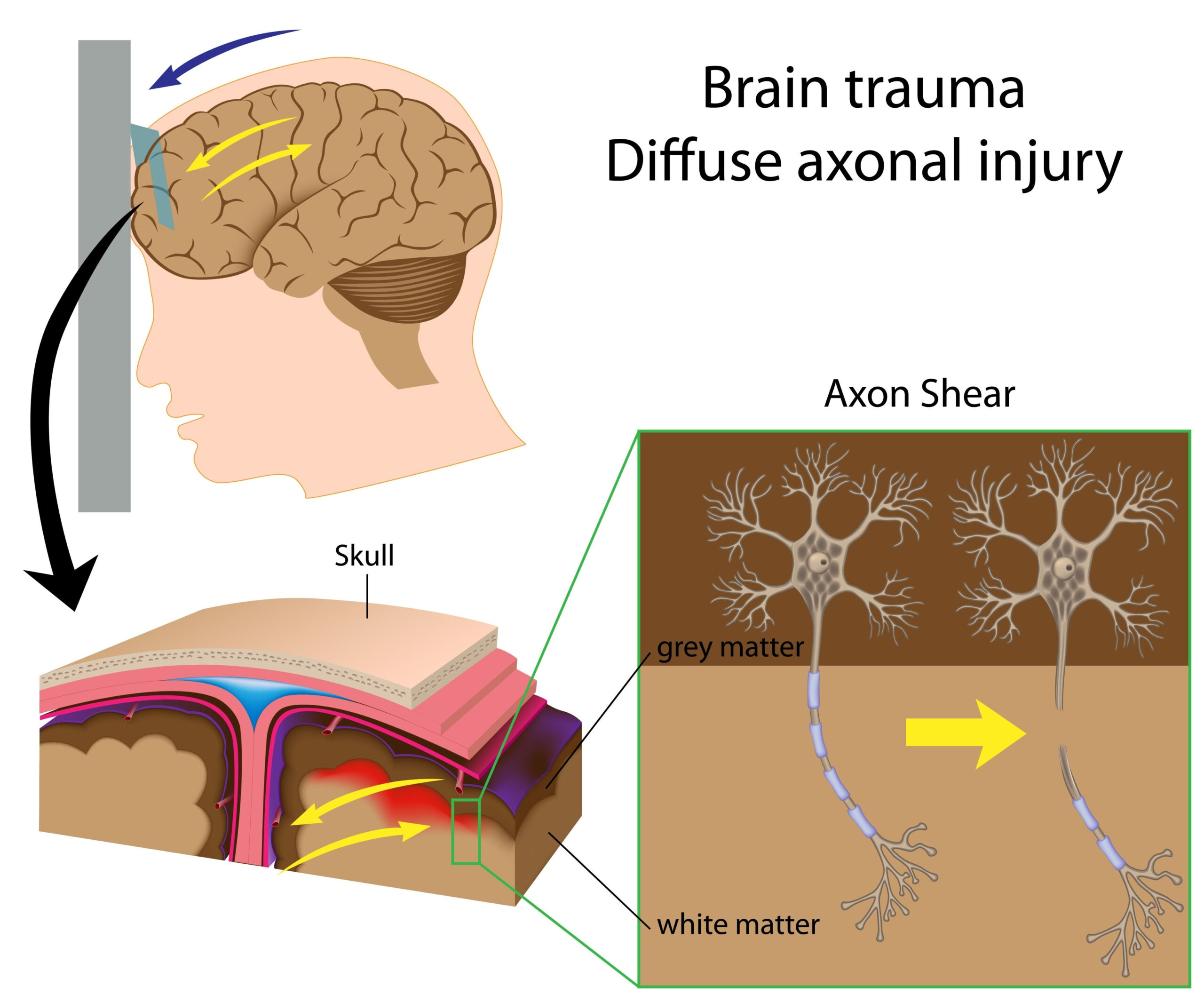
Mitochondrial dysfunction is a significant consequence of repetitive mild traumatic brain injury (mTBI). The mitochondria, often referred to as the powerhouse of the cell, are essential for energy production, particularly in high-energy demanding tissues such as the brain. Following an mTBI, alterations in mitochondrial function can impede ATP synthesis, disrupt calcium homeostasis, and trigger oxidative stress. These disturbances are exacerbated by the injury-related inflammatory response and can lead to neuronal impairment and cell death.
Research indicates that following a mild traumatic brain injury, there is a marked increase in mitochondrial fragmentation. This phenomenon, often referred to as mitochondrial fission, results in a reduced capacity for aerobic respiration. The fragmentation process is mediated by various proteins such as dynamin-related protein 1 (Drp1), which, when overactivated, leads to an imbalance in mitochondrial dynamics. A decrease in the numbers of healthy, elongated mitochondria inhibits the brain’s resilience to subsequent injuries, compounding existing vulnerabilities.
Moreover, dysfunctional mitochondria contribute to elevated levels of reactive oxygen species (ROS), resulting in oxidative stress that damages cellular components including lipids, proteins, and DNA. This oxidative damage not only affects neuronal cells but can also compromise the blood-brain barrier (BBB), further exacerbating neural injury. The BBB serves as a critical protective interface, regulating the entry of substances into the brain; its integrity is essential for maintaining homeostasis and protecting against neurotoxins.
The metabolic shift observed in energy production pathways following mTBI points to a potential metabolic crisis, as the brain tries to adapt to energy demands amidst impaired mitochondrial function. Studies show that glycolysis may become more pronounced in an effort to compensate for oxidative phosphorylation deficits, reflecting a shift away from normal energy metabolism. This metabolic remodeling could have long-term implications for brain health and function, making further investigation into these pathways essential.
Understanding the mechanisms underlying mitochondrial dysfunction following repetitive mTBI is critical for developing targeted therapeutic approaches aimed at restoring mitochondrial health and function. Research exploring mitochondrial biogenesis, antioxidant therapies, and interventions that improve mitochondrial dynamics presents exciting possibilities for mitigating the negative consequences of injury and promoting recovery in affected individuals.
Experimental Design and Techniques
To investigate the effects of repetitive mild traumatic brain injury (mTBI) on mitochondrial function and blood-brain barrier (BBB) integrity, a series of detailed experiments were conducted utilizing both in vivo and in vitro models. These studies aimed to dissect the cellular and molecular changes that occur in response to repeated milder injuries over time.
Animal models, particularly rodents, are commonly employed to simulate human brain injuries. In this context, repeated mTBI was induced using a controlled force application method, where a calibrated impact is delivered to the skull, mimicking the forces experienced in sports-related injuries or falls. This approach allows researchers to observe the cumulative effects of multiple injuries, which is critical to understanding long-term consequences on neuronal health and mitochondrial function.
Post-injury assessments typically include behavioral tests to gauge cognitive and motor function, alongside biochemical analyses to evaluate mitochondrial health and BBB permeability. For instance, researchers often measure ATP levels, mitochondrial membrane potential, and reactive oxygen species (ROS) production in brain tissues extracted post-mTBI. These parameters provide insights into mitochondrial bioenergetics and oxidative stress status following the injury.
To further analyze the integrity of the BBB, techniques such as Evans blue dye extravasation and Western blotting for tight junction proteins are employed. The use of Evans blue dye enables visualization of BBB disruption, as the dye is excluded from healthy brain tissue but can penetrate when barrier integrity is compromised. Concurrently, the expression levels of key tight junction proteins like occludin and claudin, which are crucial for maintaining BBB structure, are assessed to confirm the pathological changes observed.
In addition to in vivo studies, cellular models using primary neuronal cultures and endothelial cells are utilized to specifically dissect mitochondrial dynamics and signaling pathways. For instance, exposing these cells to injury-like conditions, such as oxygen-glucose deprivation or mechanical stress, helps in elucidating the direct effects of mTBI on mitochondrial behavior. For these experiments, techniques such as live-cell imaging and flow cytometry are employed to monitor mitochondrial fission and fusion events in real-time.
Furthermore, pharmacological interventions targeting mitochondrial function or oxidative stress responses are integrated into the experimental design. By treating injured cells with agents such as antioxidants or compounds that promote mitochondrial biogenesis, researchers can evaluate whether these therapies can mitigate the downstream effects of repeated mTBI. This approach is crucial for identifying potential therapeutic strategies that may restore mitochondrial function and protect neuronal integrity following injury.
This multifaceted experimental framework not only provides a comprehensive understanding of the biochemical alterations following repetitive mTBI but also paves the way for exploring novel therapeutic avenues focusing on mitochondrial health and BBB preservation. By employing a combination of advanced imaging techniques, molecular biology assays, and behavioral assessments, researchers are better equipped to unravel the complex interplay between injury, mitochondrial dysfunction, and neurovascular integrity.
Impact on Blood-Brain Barrier Integrity
The blood-brain barrier (BBB) serves as a fundamental protective mechanism for the central nervous system, regulating the passage of substances between the bloodstream and neuronal tissue. After repetitive mild traumatic brain injury (mTBI), this barrier often becomes compromised, leading to increased permeability that can allow potentially harmful substances to infiltrate the brain. Understanding the mechanisms behind this BBB dysfunction is vital, as it may play a crucial role in the overall pathophysiology of brain injuries.
Repetitive mTBI appears to disrupt the tightly-knit network of endothelial cells that form the BBB, primarily through the loss or dysfunction of tight junction proteins such as occludin and claudin-5. These proteins are essential in maintaining the structural integrity of the barrier, preventing the passive diffusion of various molecules. Following mTBI, studies have shown that oxidative stress and inflammation can lead to the degradation of these proteins, resulting in a breakdown of the barrier’s selectivity and allowing serum proteins and immune cells to infiltrate the brain tissue, which could further exacerbate neuronal damage.
Research using techniques such as electron microscopy has revealed alterations in the morphology of BBB endothelial cells following injury. These morphological changes, including cell swelling and retraction, contribute to a pronounced disruption in barrier function. Enhanced BBB permeability is often correlated with increased levels of inflammatory cytokines, which can further incite a cascade of tissue damage. For example, the elevation of interleukin-6 (IL-6) and tumor necrosis factor-alpha (TNF-α) in the brain after mTBI has been linked to profound alterations in BBB integrity, suggesting that inflammatory pathways are a significant contributor to barrier dysfunction.
Moreover, the role of mitochondria in BBB integrity is emerging as a critical area of study. Mitochondrial dysfunction following mTBI can lead to energy deficits, exacerbating endothelial cell stress and impairing their ability to maintain tight junction integrity. The resultant apoptotic signaling can further contribute to BBB disruption, creating a vicious cycle of injury and inflammation. Restoration of mitochondrial function has been shown in some preclinical trials to positively influence BBB recovery, highlighting the potential for therapeutic interventions aimed at mitochondrial rejuvenation.
Interestingly, the consequences of BBB dysfunction extend beyond immediate cerebral damage. With increased permeability, neurotoxic agents and inflammatory mediators can infiltrate the central nervous system, contributing to chronic neurodegenerative processes that could result in long-term cognitive deficits and emotional disturbances. This is particularly concerning in the context of repetitive injuries, where the accumulative effects of BBB breach may precipitate adverse outcomes over time, leading to conditions such as chronic traumatic encephalopathy (CTE).
Ultimately, understanding the intricate relationship between repeated mild trauma, mitochondrial dysfunction, and BBB integrity will be crucial for developing strategies aimed at preventing and treating long-term sequelae of mTBI. Investigating targeted therapies that enhance tight junction protein expression, reduce oxidative stress, and stabilize mitochondrial function presents a promising approach to safeguard BBB integrity, potentially mitigating the ill effects of repeated injuries on brain health.
Future Directions and Therapeutic Opportunities
There is a growing recognition of the need for effective therapeutic strategies to address the consequences of repetitive mild traumatic brain injury (mTBI), particularly as they relate to mitochondrial dysfunction and blood-brain barrier (BBB) integrity. While current treatment modalities remain limited, emerging research is paving the way for innovative interventions aimed at improving outcomes for individuals suffering from the effects of mTBI.
One promising avenue of therapy focuses on enhancing mitochondrial health. Mitochondrial-targeted antioxidants, such as MitoQ and SS-31, have shown potential in preclinical studies by selectively reducing oxidative stress and improving mitochondrial function. These agents work by targeting the mitochondria directly, thereby protecting against the oxidative damage caused by reactive oxygen species that accumulate following injuries. The implementation of such targeted therapies could help restore cellular bioenergetics and reduce neuronal death, which is crucial for recovery.
In addition to antioxidants, strategies that promote mitochondrial biogenesis are of considerable interest. Compounds like resveratrol and exercise have been linked to enhanced mitochondrial production, aiding in the restoration of mitochondrial function in models of brain injury. Therapeutic exercise programs, even in a controlled rehabilitation setting, may offer significant benefits not only by promoting neuroplasticity but also by augmenting mitochondrial health and improving overall cognitive and motor outcomes.
Another critical area of exploration involves modulating the inflammatory response that ensues following mTBI. Anti-inflammatory agents, particularly those targeting specific pathways, could ameliorate BBB disruption and mitochondrial impairment. For instance, inhibitors of the nuclear factor kappa-light-chain-enhancer of activated B cells (NF-κB) pathway may diminish the inflammatory cascade that leads to BBB breakdown. Furthermore, agents that selectively reduce pro-inflammatory cytokines such as IL-6 and TNF-α hold potential for preserving BBB integrity and consequently enhancing neuronal survival and function.
Additionally, regenerative medicine approaches, such as the use of stem cells, are being considered. Stem cell therapies have demonstrated the ability to differentiate into neural cell types, secrete neuroprotective factors, and promote repair processes. These therapies could provide not only cellular replenishment but also a favorable microenvironment for recovery, particularly in the context of disrupted mitochondrial function and BBB integrity.
Importantly, the development of pharmacological agents requires robust clinical translation. Clinical trials designed to evaluate the safety and efficacy of these therapeutic strategies in human populations are necessary. Biomarkers for mitochondrial dysfunction and BBB integrity would be instrumental in monitoring therapeutic responses and guiding treatment strategies. Identifying markers that indicate the extent of mitochondrial impairment or BBB disruption could allow for targeted approaches tailored to the needs of individual patients.
Ultimately, a multifaceted approach that combines mitochondrial therapies, anti-inflammatory strategies, and regenerative techniques may provide the most comprehensive benefits for individuals impacted by repetitive mTBI. By bridging the gap between laboratory findings and clinical applications, researchers and clinicians can pave the way toward improving the management of mTBI-related complications, aiming for better health outcomes and quality of life for affected individuals.