Case Presentation
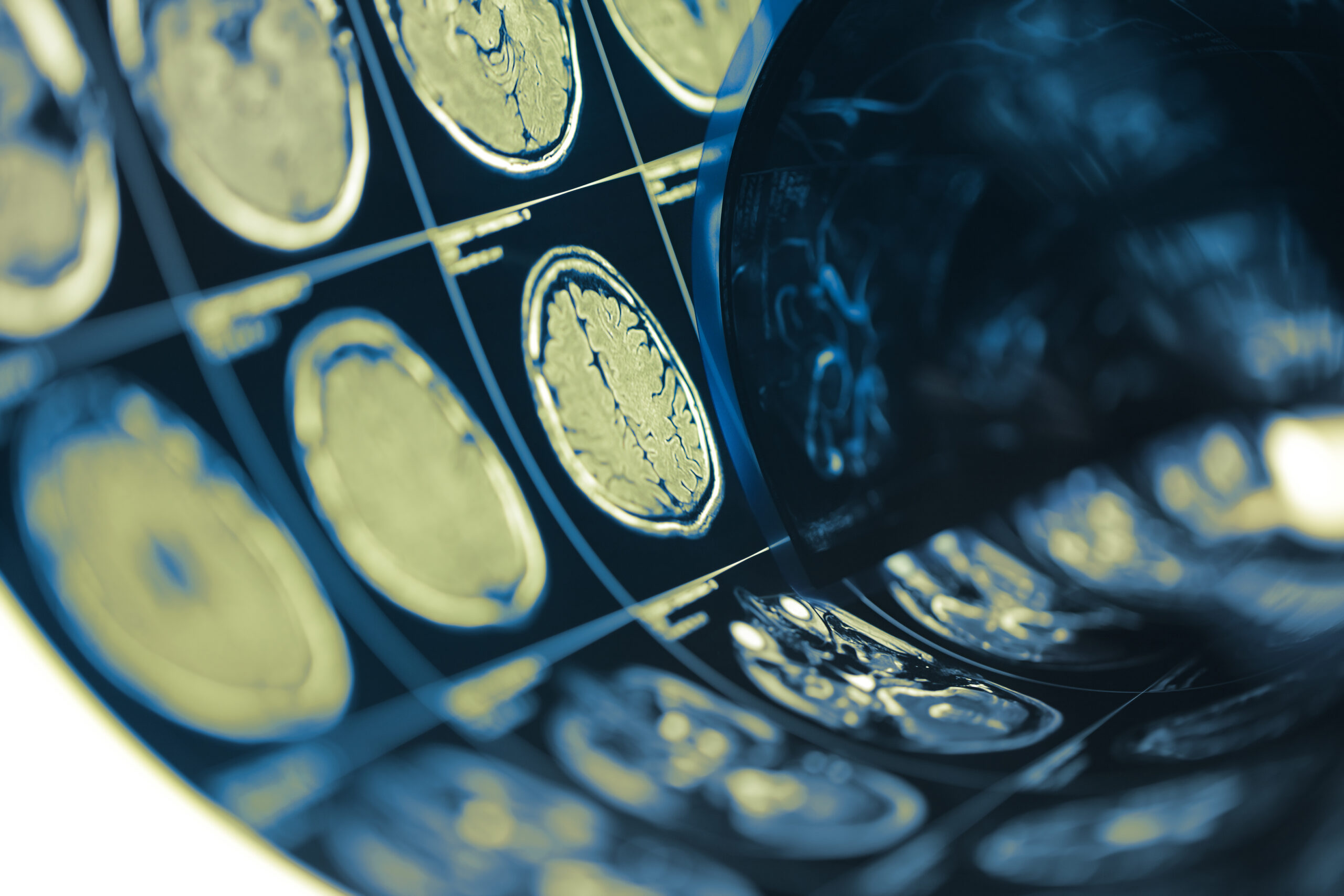
A 55-year-old female patient presented to the emergency department with chronic headaches and a recent episode of severe headache described as “the worst ever.” Upon neurologic examination, she demonstrated no focal deficits, but a magnetic resonance imaging (MRI) scan revealed an ovoid mass located in the suprasellar region. This lesion was hypointense on T1-weighted images and hyperintense on T2-weighted images, leading to the initial diagnosis of a suprasellar tumor.
Further evaluation through cerebral angiography identified a partially thrombosed anterior communicating artery (ACoA) aneurysm. The aneurysm measured approximately 9 mm in maximal diameter and was found to exert mass effect on surrounding structures, mimicking the characteristics of a neoplastic entity. The combination of these imaging findings, alongside clinical symptoms, precipitated a multidisciplinary discussion between neurosurgery and neuroradiology teams regarding the appropriate management approach for this patient.
The patient’s medical history included hypertension and hyperlipidemia, although she had no significant past surgical history or family history of aneurysms or vascular diseases. Given the potential risk of rupture associated with the aneurysm and its mimicking behavior, the decision was made to proceed with surgical intervention. Preoperative planning included discussions about the risk profile of the procedure and the expected postoperative course.
Informed consent was obtained, and the patient was prepared for a surgical approach involving aneurysmotomy and subsequent thrombectomy, aimed at addressing the aneurysm while relieving the compressive effect on the surrounding tissue. This case exemplifies the complex interplay between aneurysms and their presentations, particularly how vascular lesions can resemble tumors in imaging studies, underscoring the importance of comprehensive diagnostic approaches in neurosurgery.
Operative Techniques
The surgical intervention for the patient involved a combination of aneurysmotomy and ultrasonic thrombectomy. The procedure commenced with the patient positioned supine, and appropriate monitoring and anesthesia were established to ensure a sterile environment. A bicoronal scalp incision was made, allowing for an adequate skull exposure while minimizing potential complications from hair and soft tissue interference.
Following the incision, a craniotomy was performed to access the anterior cranial fossa. The operating microscope was utilized to enhance visualization of the intricate vascular and neural structures within this region. Once the dura mater was opened, the cerebral cortex was carefully retracted to visualize the anterior communicating artery and the aneurysm.
The partially thrombosed aneurysm was identified, measuring approximately 9 mm. It exhibited a characteristic bluish hue due to the presence of organized thrombus within the sac, which indicated both the chronic nature of the aneurysm and its potential for rupture. The technique employed for aneurysmotomy involved carefully opening the aneurysmal sac to evacuate the thrombus while preserving the integrity of the surrounding arterial structures.
Ultrasonic thrombectomy was next deployed, which utilizes high-frequency ultrasound to facilitate the fragmentation and aspiration of the thrombus. This technique is particularly advantageous as it minimizes endothelial injury compared to mechanical methods, thus preserving the vessel walls and maintaining normal blood flow. The ultrasound energy effectively disaggregates the clot, allowing for its gentle removal without damaging adjacent tissues.
Throughout the procedure, care was taken to monitor blood flow through the anterior communicating artery and its branches. This real-time assessment was pivotal in ensuring that critical vascular structures were spared during manipulation. After thorough thrombectomy, the aneurysm sac was inspected for any residual thrombus or abnormalities. Reconstruction of the aneurysm was undertaken using either an application of a graft patch or direct closure techniques as deemed appropriate, depending on the status of the aneurysmal walls post-thrombectomy.
Closure of the dura mater followed, utilizing sutures to ensure a watertight seal. The overlying bone flap was then repositioned and secured, and the scalp was sutured in layers. Careful postoperative monitoring and imaging were arranged to assess both the patient’s recovery and the status of the treated aneurysm. This careful orchestration of surgical techniques not only addressed the immediate threat posed by the aneurysm but also facilitated a resolution of the associated mass effect, thereby alleviating the patient’s neurological symptoms.
Outcome Measurements
Postoperative evaluation of the patient occurred through a combination of clinical assessments and imaging studies to ensure the successful management of the aneurysm and to monitor for potential complications. Following the surgical intervention, the patient was closely monitored in the intensive care unit (ICU) for any signs of neurological deficits or complications related to the surgery, such as hemorrhage, infection, or vasospasm.
The patient’s neurological status was assessed daily through standardized examinations, evaluating factors such as level of consciousness, motor and sensory functions, and any cranial nerve deficits. Attention was placed on identifying any changes in the patient’s baseline neurological status that could indicate complications. With the surgical approach designed to alleviate mass effect and prevent aneurysmal rupture, the clinical focus was on ensuring the patient’s recovery trajectory remained positive.
Imaging follow-ups were conducted using MRI and CT angiography at intervals of 1 week, 3 months, and 6 months postoperatively. Initial imaging confirmed the absence of residual thrombus within the aneurysmal sac and assessed the patency of the anterior communicating artery and adjacent vascular structures. These imaging modalities are critical for ensuring the complete resolution of the previous mass effect and for detecting any early signs of possible complications such as postoperative bleeding around the surgical site or new vascular changes.
The measurements of clinical outcomes were assessed using specific scales, including the Glasgow Coma Scale (GCS) and the modified Rankin Scale (mRS), which help quantify the patient’s functional status and level of independence after surgery. These scales provided an objective means to evaluate the impact of the surgical intervention on her overall well-being. A favorable outcome was defined as a reduction in the severity of headaches and an improvement in neurological function, aiming for a score of mRS of 0-2, indicating little to no disability.
Additionally, patient-reported outcomes were collected, allowing an assessment of her quality of life post-surgery. This included surveys that addressed symptoms such as headaches, cognitive function, and emotional well-being. The goal was to evaluate not only the physiological implications of aneurysm treatment but also to consider the psychological and social aspects of recovery.
The overall success of the surgical intervention was determined not only by the immediate postoperative outcomes but also by the long-term maintenance of the patient’s neurological status and absence of aneurysmal recurrence. Regular follow-up visits and imaging played a crucial role in establishing a comprehensive outcome measurement framework, promoting adjustments to post-surgical care as necessary based on the patient’s evolving condition and recovery progress.
These outcome measurements align with current standards in neurovascular surgery, emphasizing the importance of continuous evaluation of surgical efficacy and patient safety to optimize rehabilitation and recovery protocols in future cases.
Future Considerations
As advancements in neurovascular surgery continue to evolve, several factors must be taken into account for optimizing surgical techniques and patient outcomes in cases involving partially thrombosed aneurysms. One significant area of consideration is the integration of novel imaging modalities and their potential to enhance preoperative planning and intraoperative navigation. The use of advanced imaging, such as intraoperative MRI or digital subtraction angiography, could provide real-time feedback on the aneurysm’s morphology and blood flow dynamics, allowing surgeons to make more informed decisions during the procedure.
Moreover, research into the long-term effectiveness of various aneurysm treatments is crucial. Given that some aneurysms may demonstrate a propensity for regrowth or new formation post-treatment, systematic follow-up studies assessing the long-term durability of surgical options will be beneficial. This includes evaluating the comparative effectiveness of surgical intervention against endovascular techniques, particularly for complex aneurysm presentations. Understanding the relative risks and benefits of these approaches could lead to tailored treatment plans that maximize patient safety and efficacy.
Furthermore, ongoing studies aimed at understanding the genetic and environmental risk factors contributing to aneurysm development may elucidate preventive strategies. Identifying individuals at high risk due to hereditary patterns or predisposing conditions is essential. Implementing systematic screening protocols, particularly for populations with a familial history of cerebral aneurysms, could facilitate early intervention and potentially mitigate the risks of rupture.
Educational initiatives targeting both patients and healthcare providers are essential to improve awareness and understanding of aneurysms. Ensuring that patients are informed about the signs and symptoms correlating with aneurysm complications can prompt timely medical consultations and evaluations. Additionally, enhancing the knowledge base of clinicians regarding emerging treatment strategies and current guidelines will facilitate better care decisions in rapidly changing clinical landscapes.
Finally, rigorous clinical trials exploring new therapeutic agents or innovative surgical techniques should be prioritized. The adoption of adjunctive therapies, such as pharmacological agents that may impact aneurysm stability or promote vascular healing, represents a promising avenue. Rigorous evaluation of these therapies, combined with traditional surgical approaches, could enhance overall treatment paradigms and improve patient prognoses.
An integrated approach combining advanced imaging techniques, long-term outcome assessments, genetic profiling, enhanced patient education, and innovative therapeutic research will be paramount in shaping the future of aneurysm management and ensuring optimal patient care in complex neurovascular scenarios.