Background and Rationale
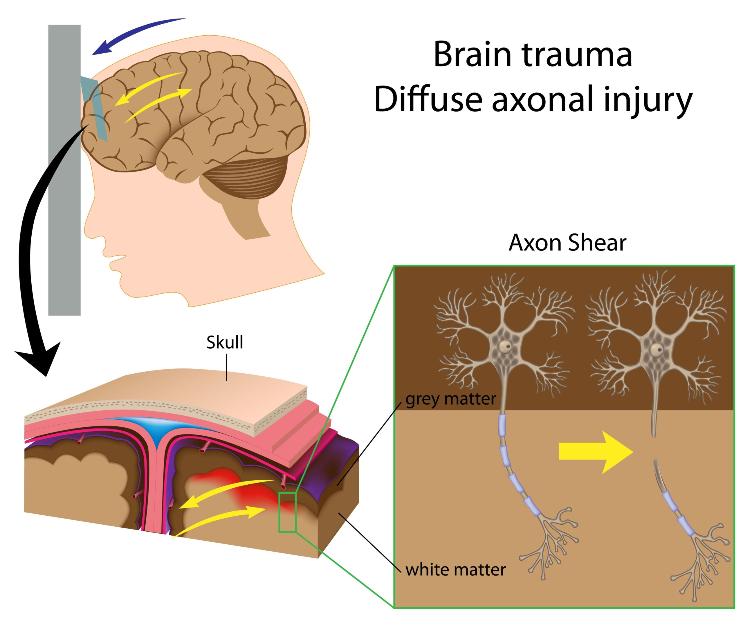
Acute posttraumatic headache (aPTH) is a common and distressing condition that often arises following traumatic brain injury (TBI). This phenomenon significantly impacts the quality of life in patients, leading to considerable healthcare burdens. Understanding the underlying neurobiological mechanisms that contribute to aPTH is essential for developing targeted intervention strategies.
Research indicates that the neural networks involved in pain processing may become altered in the aftermath of a TBI. Specifically, changes in functional connectivity within certain brain regions associated with pain perception and modulation may predispose individuals to experience persistent headaches. Emerging studies have harnessed advanced imaging techniques, such as functional magnetic resonance imaging (fMRI), to explore these alterations in greater detail, providing insights into how connectivity differences correlate with clinical symptoms.
The rationale for investigating functional connectivity in patients with aPTH lies in the observation that traditional pain assessment methods often fail to encapsulate the full experience of chronic headache sufferers. Quantifying the brain’s connectivity patterns may reveal hidden relationships between specific neural pathways and headache severity, thus allowing for more precise therapeutic approaches.
Furthermore, understanding how brain network alterations manifest after trauma could illuminate potential biomarkers for aPTH, facilitating earlier diagnosis and personalized treatment plans. Given the complex interplay of biological, psychological, and environmental factors at play, this line of inquiry is crucial in enhancing therapeutic outcomes and coping strategies for those affected by this condition.
In summary, addressing the functional connectivity alterations associated with acute posttraumatic headache is imperative not only for scientific enrichment but also for improving patient care in populations grappling with the sequelae of traumatic brain injuries.
Study Design and Participants
To investigate the functional connectivity alterations associated with acute posttraumatic headache (aPTH), a cross-sectional study design was employed. This approach enables a snapshot analysis of individuals with varying degrees of headache post-trauma, allowing researchers to identify patterns and associations pertinent to neural connectivity in real-time.
Participants were recruited from neurology clinics with expertise in traumatic brain injury management. The selection criteria included adults aged 18 to 65 who had sustained a mild to moderate TBI and developed aPTH within the first month following the injury. This time frame is critical, as it allows for the observation of acute phase alterations before chronic headache patterns potentially establish.
Exclusion criteria were meticulously defined to eliminate confounding variables. Individuals with a history of chronic headaches prior to the injury, a previous history of neurological disorders, or those currently taking medications known to affect central nervous system functioning were not included. This careful selection process aimed to ensure that the observed alterations in functional connectivity could confidently be attributed to the recent traumatic event.
Following the recruitment process, participants underwent comprehensive clinical assessments. These assessments included standardized headache evaluations based on the International Classification of Headache Disorders criteria, ensuring a consistent and accurate diagnosis of aPTH. Patient-reported outcome measures were utilized to quantify headache severity, frequency, and the associated impact on daily living activities.
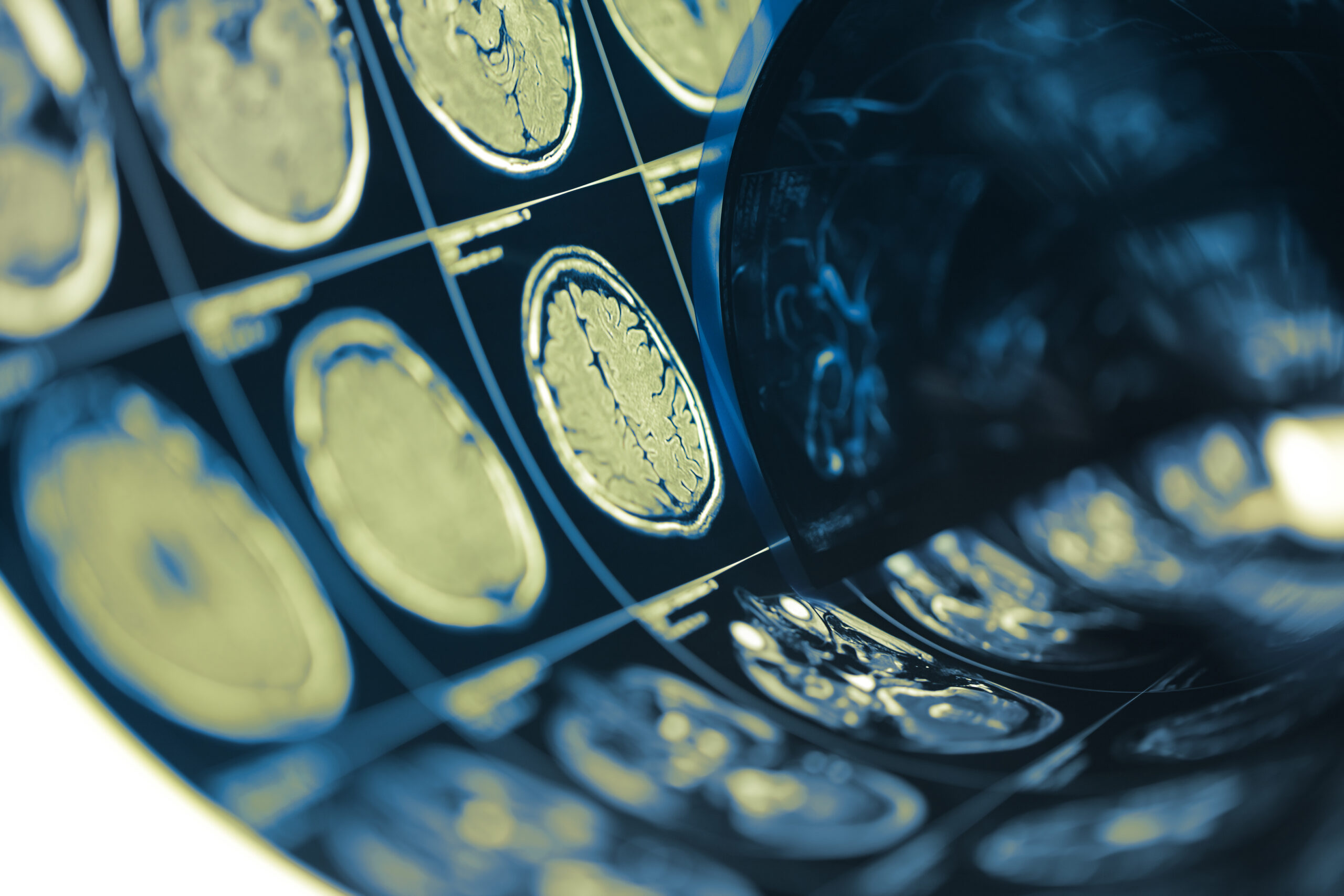
Neuroimaging was conducted using functional magnetic resonance imaging (fMRI) to assess brain activity and connectivity. During the fMRI session, participants were instructed to rest with their eyes closed, allowing for the measurement of intrinsic brain activity without the interference of external stimuli. This resting-state fMRI methodology is instrumental in capturing the baseline connectivity patterns of different neural networks, particularly those implicated in pain processing.
Moreover, advanced analytical techniques were applied to the fMRI data. Seed-based correlation analysis was utilized to identify connectivity between predefined regions of interest, focusing on areas known to play roles in nociception and pain modulation, such as the anterior cingulate cortex, insula, and thalamus. This approach enabled researchers to map out alterations in connectivity that may correlate with the clinical presentation of aPTH.
Demographic data, including age, sex, and education level, were collected to explore potential modifiers in the analysis. Understanding variations in brain connectivity based on demographic factors can provide further insights into the complexities of posttraumatic headache syndromes.
In summary, this study’s design, including detailed participant selection and robust neuroimaging analyses, establishes a comprehensive framework for understanding the neural underpinnings of acute posttraumatic headache. Through this work, researchers aim to unveil the specific connectivity alterations that contribute to the painful experiences of individuals recovering from traumatic brain injuries.
Results and Analysis
The findings from the functional magnetic resonance imaging (fMRI) assessments revealed distinct alterations in brain network connectivity in individuals suffering from acute posttraumatic headache (aPTH). Notably, there was a significant disruption in the functional connectivity between regions traditionally associated with pain processing, such as the anterior cingulate cortex, insula, and thalamus. These changes suggest that the neural mechanisms involved in pain modulation may be compromised following trauma.
The seed-based correlation analysis indicated a marked decrease in connectivity strength between the anterior cingulate cortex and the insula in patients with aPTH compared to those without headache post-injury. This dysfunction in connectivity could elucidate why individuals with aPTH experience intensified pain perception and related symptoms, as these brain areas are critical for integrating sensory and emotional aspects of pain. In contrast, the connectivity strength between these regions and the thalamus showed variability, indicating a more complex relationship depending on individual differences in pain experiences.
Further analysis of demographic data highlighted that variables such as age and sex influenced the identified connectivity patterns. For instance, younger patients exhibited greater alterations in connectivity compared to older cohorts, suggesting that developmental factors may play a role in how neural networks respond to trauma. Additionally, female participants demonstrated higher connectivity disruption, potentially reflecting gender differences in pain sensitivity and coping mechanisms.
Clinical correlations were drawn between the observed connectivity changes and the severity of headache symptoms reported by participants. A positive correlation was found between the degree of connectivity loss in the anterior cingulate cortex and the intensity of headache experienced, emphasizing the relationship between altered brain function and clinical manifestations of aPTH. Interestingly, frequency of headaches also correlated with changes in connectivity patterns, underscoring the potential for these alterations to predict headache recurrence in affected individuals.
Advanced statistical methods, including machine learning algorithms, were employed to evaluate the data further. These methods allowed researchers to classify participants based on their connectivity profiles, effectively distinguishing between individuals who developed persistent aPTH and those who demonstrated resolution of symptoms. Such predictive models could serve as a powerful tool for early intervention strategies, tailoring treatments to those at higher risk of chronic headache development following TBI.
Overall, the results underscore the intricate connection between altered brain network functionality and the clinical presentations of acute posttraumatic headache. These insights not only deepen our understanding of the neurobiological underpinnings of aPTH but also pave the way for future research exploring targeted therapeutic modalities. By elucidating the specific neural circuitry involved, clinicians can better diagnose and treat headache disorders resulting from traumatic brain injuries.
Future Directions
The exploration of functional connectivity alterations in acute posttraumatic headache (aPTH) opens numerous avenues for future research endeavors aimed at unraveling the complexities of this condition. An essential next step involves longitudinal studies that track connectivity changes over time in individuals after traumatic brain injury (TBI). Such studies could elucidate how brain network adaptations evolve from the acute phase of aPTH into potential chronic pain states, identifying critical periods for intervention. Indicators of resilience or vulnerability could be established, enabling healthcare providers to implement early preventative strategies in patients at higher risk.
Moreover, there is a pressing need to integrate multimodal imaging approaches alongside functional magnetic resonance imaging (fMRI). Combining fMRI with techniques such as diffusion tensor imaging (DTI) may provide a more comprehensive understanding of the structural connectivity and microstructural changes that accompany alterations in functional connectivity. This multimodal perspective could offer insights into how disruptions in white matter integrity influence the neural circuitry associated with pain perception, thereby enhancing our understanding of the pathological basis of aPTH.
Investigating the impact of different therapeutic modalities on functional connectivity is another promising direction. Interventions such as neurofeedback, cognitive-behavioral therapy, and pharmacological treatments may modulate brain connectivity and provide practical applications in clinical settings. Studies assessing how these interventions alter functional connectivity patterns can guide the development of tailored, evidence-based treatment options that target the distinct neurobiological underpinnings of aPTH.
Furthermore, exploring the relationship between psychosocial factors and brain connectivity could enrich the narrative surrounding aPTH. Factors such as anxiety, depression, and trauma history may interact with the neural processes underlying headache symptoms. Comprehensive assessments that include psychological evaluations alongside imaging studies would provide a broader context for understanding how these elements influence pain pathways and treatment outcomes.
Finally, the pursuit of potential biomarkers derived from neuroimaging data represents an exciting frontier. By identifying specific connectivity profiles associated with headache severity or chronicity, researchers could pave the way for objective diagnostic criteria. This objective measurement could revolutionize the clinical approach to diagnosing and managing aPTH, moving away from subjective self-reports toward a more precise, data-driven methodology.
In conclusion, these future directions hold the potential to significantly advance our understanding of acute posttraumatic headache. Integrating novel imaging techniques, longitudinal analyses, therapeutic interventions, and psychological evaluations, while also exploring biomarkers, are critical steps in unraveling the intricate relationship between brain function and headache pathophysiology. By continually refining and expanding the scope of research in this field, we can aim to improve diagnostic accuracy and therapeutic effectiveness for individuals affected by this debilitating condition.