Study Overview
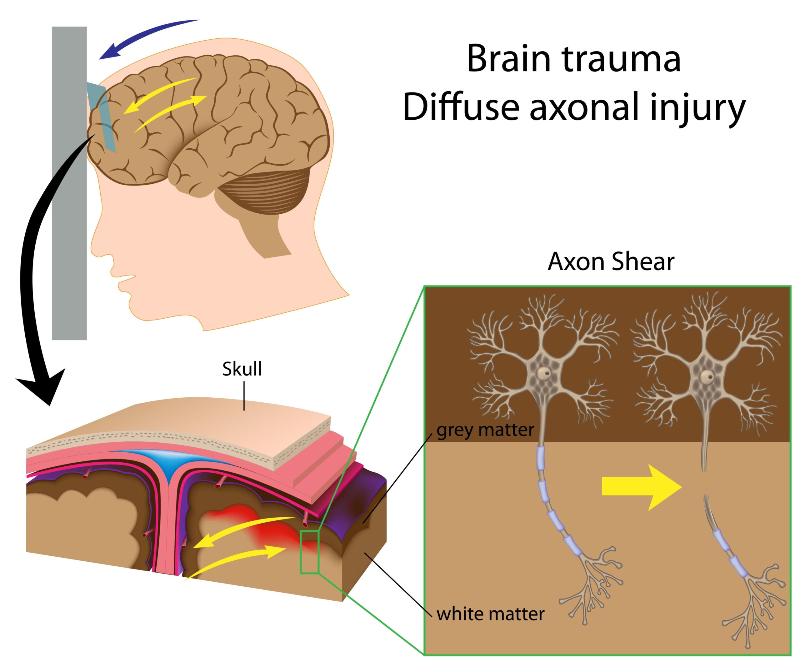
This research investigates the impact of repeated exposure to low-level blast waves on brain structure, specifically focusing on military personnel such as breachers and snipers. These individuals often operate in environments where blast exposure is common, raising concerns about potential long-term effects on their neurological health. The study aims to elucidate how this type of blast exposure is linked to region-specific reductions in gray matter volume, which could provide insights into the physiological consequences of such exposures.
The investigation is grounded in the premise that low-level blasts, while not producing immediate traumatic injuries, may still exert significant cumulative effects on cognitive functioning and brain integrity over time. Previous studies have documented a relationship between high-intensity blast exposure and brain injuries; however, the current focus is on lower-level exposures, which are often overlooked. As a result, this study contributes to a growing body of literature examining subtle yet critical changes in brain morphology as the consequence of repetitive, non-penetrating blast events.
In conducting the study, researchers utilized advanced neuroimaging techniques to assess and measure gray matter volume in specific brain regions thought to be affected by blast exposure. By comparing these measurements across different groups—those frequently exposed to low-level blasts versus a control group—researchers aimed to identify any significant differences in brain structure associated with military duties.
Ultimately, this inquiry is essential for understanding the long-term neurological impacts of military service, highlighting the need for further investigation into preventive measures and interventions that may mitigate brain damage associated with such exposures.
Methodology
The study employed a robust methodological framework to evaluate the effects of low-level blast exposure on brain structure among military personnel. Participants were divided into two primary groups: individuals engaged in roles with significant exposure to low-level blasts, such as breachers and snipers, and a control group consisting of personnel with minimal or no blast exposure. This comparative approach is critical for isolating the effects of blast exposure on brain morphology.
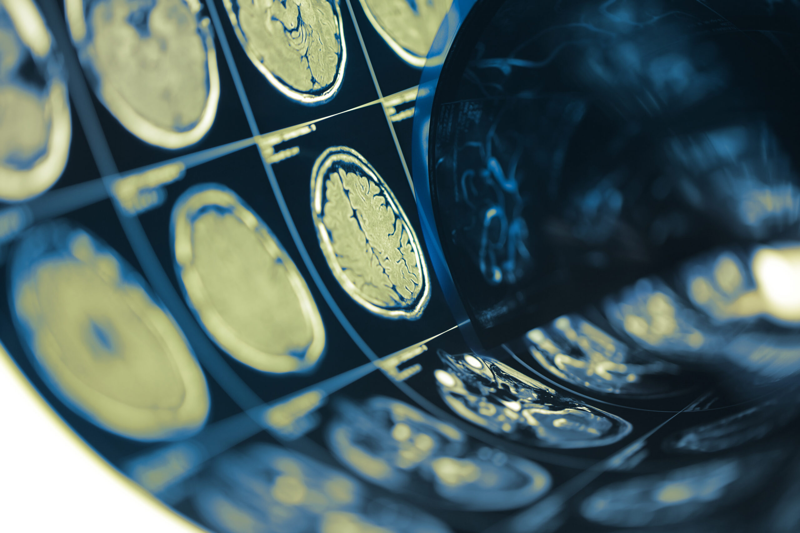
To gauge the impact of exposure, researchers utilized neuroimaging techniques, particularly high-resolution magnetic resonance imaging (MRI), to quantify gray matter volume in specific brain regions associated with cognitive function, emotional regulation, and sensory processing. These regions included the prefrontal cortex, hippocampus, and amygdala, all of which are vital for higher-order cognitive processes and emotional responses.
The neuroimaging procedures were conducted under optimal conditions to ensure accuracy and reliability. Participants underwent multiple scans to account for variability and to achieve reproducibility in results. To facilitate a comprehensive analysis, specific regions of interest (ROIs) were identified based on prior literature suggesting vulnerability to structural changes linked with blast exposure.
In conjunction with neuroimaging, the study implemented a detailed survey to assess participants’ history of military service, including duration and intensity of exposure to blast environments. This survey included standardized psychological assessments to evaluate potential cognitive and emotional effects resulting from their military experiences.
Statistical analysis of the neuroimaging data incorporated advanced imaging processing software, which allowed for the measurement of gray matter volume in a standardized manner. Researchers applied voxel-based morphometry (VBM), enabling the identification of regional brain differences while controlling for potential confounding variables, such as age, sex, and educational background.
The combination of these methodologies not only provided a clear framework for assessing the impact of low-level blast exposure on brain structure but also allowed researchers to establish correlations between structural changes and cognitive/emotional assessments, drawing a more comprehensive picture of the possible consequences of military duty under such conditions.
Key Findings
The results of this study provide compelling evidence of the subtle yet significant impacts of low-level blast exposure on brain structure among military personnel, particularly breachers and snipers. The data revealed that individuals with repeated exposure to blast waves exhibited localized reductions in gray matter volume in specific regions of the brain, notably the prefrontal cortex and hippocampus. These findings align with the hypothesis that even low-intensity blast events can exert cumulative effects on neural integrity over time.
In particular, reductions in gray matter in the prefrontal cortex, which plays a crucial role in decision-making, impulse control, and social behavior, may have profound implications for cognitive functioning. Similarly, changes in the hippocampus, a region essential for memory formation and spatial navigation, could potentially affect both learning and retraining capabilities. The inclusion of the amygdala, involved in emotional processing, underscores the possibility that repeated blast exposure may also disrupt emotional regulation, further complicating the psychological profile of affected personnel.
The statistical analysis confirmed that these reductions were statistically significant when comparing the blast-exposed group with the control group, indicating that the differences were not merely due to chance or external variables. Furthermore, correlations between the extent of gray matter loss and performance on cognitive assessments highlighted a possible link between structural changes and functional deficits, suggesting that the impact of blast exposure goes beyond structural integrity, affecting operational readiness and mental health outcomes.
Interestingly, the study also revealed that the degree of gray matter reduction varied according to the intensity and duration of blast exposure, suggesting a potential dose-response relationship. This insight emphasizes the need for ongoing monitoring and evaluation of personnel frequently exposed to blast environments, as cumulative exposure may increase the risk of long-term neurological consequences.
These findings illuminate the hidden vulnerabilities associated with military service in high-blast environments. By identifying specific brain regions that are negatively impacted, the research lays the groundwork for future studies aimed at developing targeted interventions and rehabilitation strategies to support the cognitive and emotional well-being of military personnel affected by such exposures.
Clinical Implications
The findings of this study highlight critical clinical implications for military personnel repeatedly exposed to low-level blast waves, particularly breachers and snipers. The documented reductions in gray matter volume within specific brain regions underline the urgent need for enhanced monitoring and assessment protocols for individuals in high-risk roles. Understanding that even low-intensity blasts can lead to significant structural changes in the brain is vital for developing preventive strategies aimed at safeguarding the cognitive health of military personnel.
Healthcare providers and military psychologists should be aware of the potential long-term cognitive and emotional consequences associated with low-level blast exposure. Notably, the involvement of the prefrontal cortex suggests that affected individuals may experience challenges in decision-making, impulse control, and social interactions, which could directly impact their operational effectiveness in military settings. Furthermore, the changes observed in the hippocampus could lead to difficulties with memory and learning, essential skills for training and mission success.
As the study indicates a potential dose-response relationship between blast exposure and gray matter alteration, military health programs could benefit from implementing routine neurocognitive evaluations for service members in blast-prone environments. These assessments could help identify those at risk of developing cognitive impairments, allowing for early intervention tailored to individual needs. Such interventions might include cognitive training exercises, psychological support, and strategies designed to enhance coping mechanisms and resilience against stressors associated with military duties.
Moreover, the research underscores the importance of developing protective measures within military training and operational protocols. The findings could prompt military organizations to reevaluate exposure limits to low-level blasts and consider implementing protective equipment or modified training methods that could minimize exposure to potentially harmful blast waves. While traditional concerns have focused on acute traumatic brain injuries, this study suggests a paradigm shift towards recognizing the cumulative impact of low-level exposure, thereby advocating for a comprehensive approach to brain health.
In light of the increasing awareness surrounding mental health in military populations, integrating these findings into training and health guidance will be essential. Enhanced educational initiatives aimed at service members can help them understand the risks associated with repeated blast exposure and empower them to advocate for their health proactively.
Ultimately, addressing the implications of this research may not only improve individual health outcomes but also enhance overall military performance by ensuring that personnel are mentally fit and cognitively resilient in high-stakes environments. Moreover, as our understanding of brain health continues to evolve, the insights gained from this study will be pivotal in informing future research directions and clinical practices aimed at supporting the well-being of military personnel exposed to low-level blasts.