Study Overview
The investigation into intracranial brain deformation involves analyzing post-mortem specimens of human brains subjected to various impact scenarios. This study aims to understand how different forces can alter the structure of the brain, which has implications for both medical research and legal contexts, especially in the evaluation of traumatic brain injuries (TBIs). By examining specimens from individuals who have experienced different levels of impact, the researchers aspire to reveal the biomechanical responses of the brain tissue.
The choice of utilizing post-mortem specimens permits researchers to conduct extensive assessments on the brain’s physical state after death, offering insights that are often unattainable through live imaging techniques. This retrospective approach is essential for correlating observed tissue deformation with specific types of impact, which can range from blunt force trauma to more complex acceleration-deceleration events, mimicking those experienced in vehicular accidents or falls.
The study emphasizes the relationship between the magnitude and direction of applied forces and the resultant physiological changes in brain composition. Through careful experimentation and analysis, researchers aim to enhance the existing knowledge surrounding brain injuries, thus providing crucial information that may inform future developments in both therapeutic strategies and preventive measures for TBIs. The findings are expected to contribute significantly to the current body of literature in neurotrauma, aiding in the development of improved diagnostic techniques and potential avenues for treatment.
This examination is particularly relevant given the increasing recognition of TBIs as a major public health concern, prompting the need for comprehensive studies that elucidate the mechanics of brain injury. With a focus on scientific rigor, the study employs advanced imaging and modeling techniques to provide a detailed understanding of intracranial dynamics under various impact conditions, setting the stage for further exploration in this critical area of research.
Methodology
The methodology of this study was designed to comprehensively examine the impacts of various forces on intracranial brain deformation using a systematic approach. The research employed a well-defined protocol for sample selection, preparation, and analysis, ensuring that the results would be both reliable and relevant.
Post-mortem human brain specimens were procured from a controlled cohort that included individuals with a range of demographic characteristics, such as age, sex, and health status prior to death. This variation facilitated a comparative analysis across different tissue types and conditions. Specimens were selected based on their availability and the circumstances of death, ensuring a diverse representation of impact scenarios.
To simulate the mechanical forces that the brain experiences during traumatic events, a series of dynamic impact tests were conducted. Each specimen was subjected to a predefined set of impact conditions that mimicked real-world situations, including both blunt force and acceleration-deceleration events. These conditions were critical for examining the resulting biomechanical changes within the brain tissue. The impact apparatus was calibrated to deliver varying magnitudes of force, allowing researchers to collect data on how the brain deforms under different circumstances.
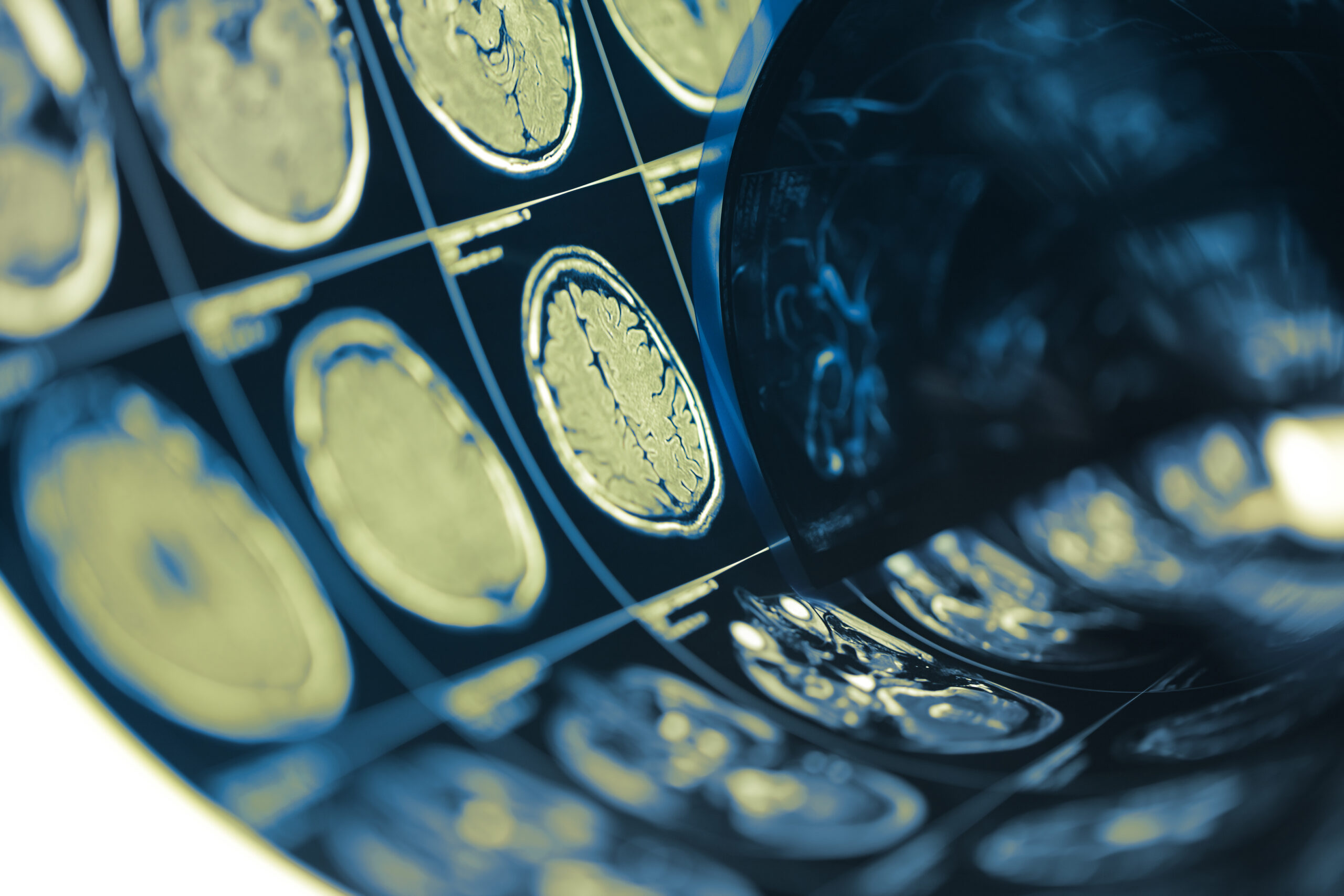
The deformation of the brain was quantitatively assessed using advanced imaging techniques, including magnetic resonance imaging (MRI) and high-resolution computed tomography (CT) scans. These imaging methods provided detailed visualizations of the brain’s structural integrity before and after impact exposure, enabling researchers to identify specific areas of deformation and analyze the extent of damage. Additionally, three-dimensional (3D) modeling software was utilized to create accurate representations of brain anatomy, enhancing the precision of the deformation measurements.
Following imaging, the data was analyzed using sophisticated computational models designed to interpret the biomechanical responses of brain tissue. Finite element analysis (FEA) techniques were employed to simulate and predict how different types of impact forces could lead to specific patterns of deformation, allowing researchers to establish correlations between force vectors and observed tissue responses.
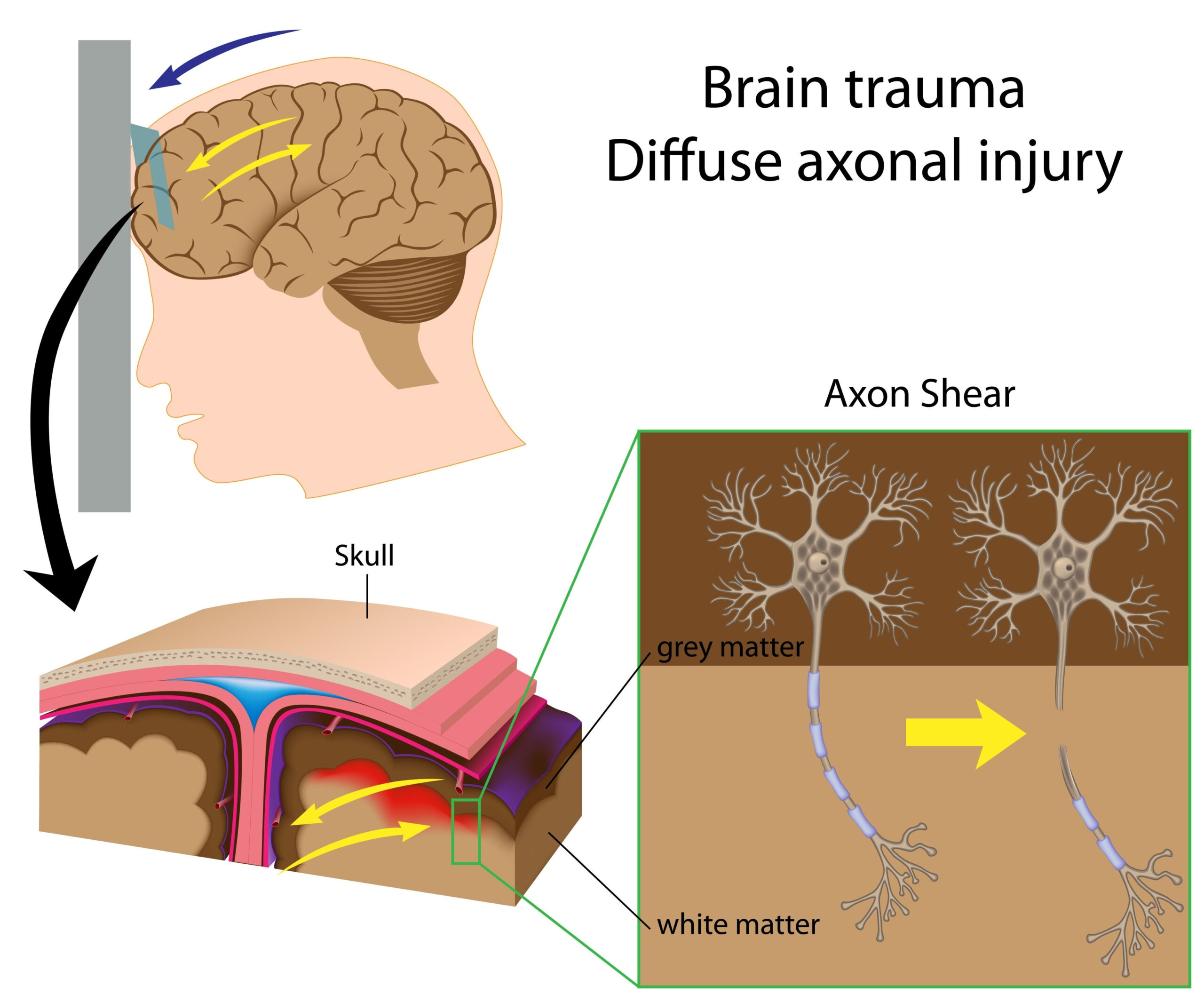
Furthermore, a series of post-mortem examinations were performed on specimens that demonstrated significant deformation. Histological analyses were conducted to assess microstructural changes within the brain tissue. This involved carefully examining the cellular architecture to identify any pathological features resulting from the impact events, such as axonal injury or glial activation.
The data collected from both the imaging and histological analyses were integrated into a comprehensive dataset. Statistical methods were utilized to determine the significance of findings, providing a robust framework for interpreting the results. This combination of dynamic testing, advanced imaging, modeling techniques, and histological evaluation forms a multi-faceted methodology that aimed to unlock deeper insights into intracranial dynamics and trauma mechanisms. Ultimately, this rigorous methodological approach allowed for a thorough investigation of the complex interactions between impact forces and brain deformation, paving the way for potential advancements in understanding and addressing traumatic brain injuries.
Key Findings
The analysis of intracranial deformation in post-mortem human specimens revealed several significant insights into how the brain responds to varying impact conditions. Primarily, the data indicated a clear correlation between the magnitude of the applied force and the resulting degree of brain deformation. Specimens subjected to high-energy impacts exhibited more extensive and localized deformation compared to those exposed to lower-energy forces. This finding underscores the importance of force intensity in determining the extent of trauma and the consequent structural changes within the brain.
Additionally, the directionality of the impact forces played a crucial role in shaping the deformation patterns observed. Blunt forces applied at perpendicular angles to the skull resulted in distinct morphological changes compared to oblique impacts, which elicited different responses, particularly in the cortical regions. The study found that localized deformation was often accompanied by widespread damage to surrounding neural structures, suggesting that even impacts that appear focused can have far-reaching effects on brain integrity.
Advanced imaging analyses, specifically through the utilization of MRI and CT scans, delineated various regions of vulnerability within the brain. Areas such as the frontal and temporal lobes demonstrated increased susceptibility to deformation and injury, consistent with observations from clinical cases of traumatic brain injuries. The spatial distribution of damage in the specimens corroborates findings from in vivo studies, establishing a link between post-mortem analyses and real-world scenarios.
Quantitative assessments through finite element analysis illustrated specific deformation models relating to different types of trauma. The simulations provided predictive insights into how varying intensities and angles of impact lead to distinctive deformation trajectories, reinforcing the biomechanical principles that govern brain injury. These results contribute to the development of injury criteria that may improve the understanding of head trauma in living subjects, particularly in contexts such as sports or vehicular collisions.
Histological evaluations further reinforced the biomechanical findings, revealing microstructural consequences of the mechanical forces. Notably, axonal injury was prevalent in areas where deformation was most severe. The presence of glial activation and inflammatory markers indicated secondary injury processes, which are critical in the progression of traumatic brain injuries. This highlights a complex interplay between mechanical forces and biological responses that could influence recovery trajectories following injury.
Overall, the findings of this study hold significant implications for developing future research avenues and clinical applications. By elucidating the biomechanical pathways and resultant structural changes within the brain, there is potential to enhance existing diagnostic frameworks and therapeutic strategies targeting traumatic brain injuries. The results not only add depth to the understanding of intracranial dynamics but also emphasize the need for continued exploration into protective measures, treatment modalities, and the long-term effects of brain trauma.
Strengths and Limitations
The strengths of this study are rooted in its comprehensive approach to understanding the mechanical forces that influence brain deformation. Utilizing well-defined methodologies, the research capitalizes on advanced technologies, including high-resolution imaging and sophisticated modeling techniques. This integration enhances the reliability of the findings by providing a robust framework for analyzing the biomechanical responses of human brain specimens to trauma. The use of post-mortem specimens allows for an unprecedented examination of brain deformation, offering an opportunity to correlate structural changes with specific impact conditions in ways that are not possible with live subjects.
One major strength lies in the diverse cohort of post-mortem subjects, encompassing a range of demographics including varied age, sex, and health statuses. This diversity is crucial as it allows researchers to explore how these factors may influence brain response to trauma. Moreover, the simulated impact conditions mirror real-world scenarios, enhancing the ecological validity of the findings. The study’s use of quantitative measures of deformation, along with histological analyses, provides a multi-layered understanding of the mechanical and biological impacts of brain injuries.
However, several limitations should be acknowledged. One significant constraint is the inherent variability in individual brain anatomy, which could affect the generalizability of findings. While the study aims to establish correlations between impact conditions and deformation, individual differences in brain structure and cerebrospinal fluid dynamics can lead to divergent responses to similar forces. This variability can complicate the translation of results from post-mortem specimens to living individuals.
Another limitation is the focus on post-mortem analysis, which, despite its advantages, does not account for the dynamic processes that occur during life. Significant aspects of brain injury, such as immediate neurochemical responses, inflammatory reactions, and the body’s systemic reactions to trauma, can only be partially inferred from static analyses. As such, while the structural insights are invaluable, the full spectrum of injury dynamics in live patients cannot be captured.
Additionally, the use of a limited number of specimens may restrict the statistical power of the findings, potentially diminishing the ability to detect subtle but clinically significant effects. Future studies could benefit from larger sample sizes and more diverse types of impacts to validate and expand upon these findings.
Moreover, while finite element analysis can provide predictive modeling of brain deformation, the accuracy of these predictions hinges on the precise calibration of impact conditions and the material properties of brain tissue. Any discrepancies in these parameters may affect model outcomes and interpretations. Continued refinement of computational models and validation against clinical data will be essential for enhancing the predictive accuracy of biomechanical simulations.
In summary, while the study offers substantial insights into intracranial brain deformation under varying impact forces, a careful consideration of its limitations is necessary. Recognizing the complexities of biological responses to trauma will guide further research in this field, ensuring a more nuanced understanding of traumatic brain injuries and their implications for patient outcomes.