Brain Changes in Blast-Exposed Soldiers
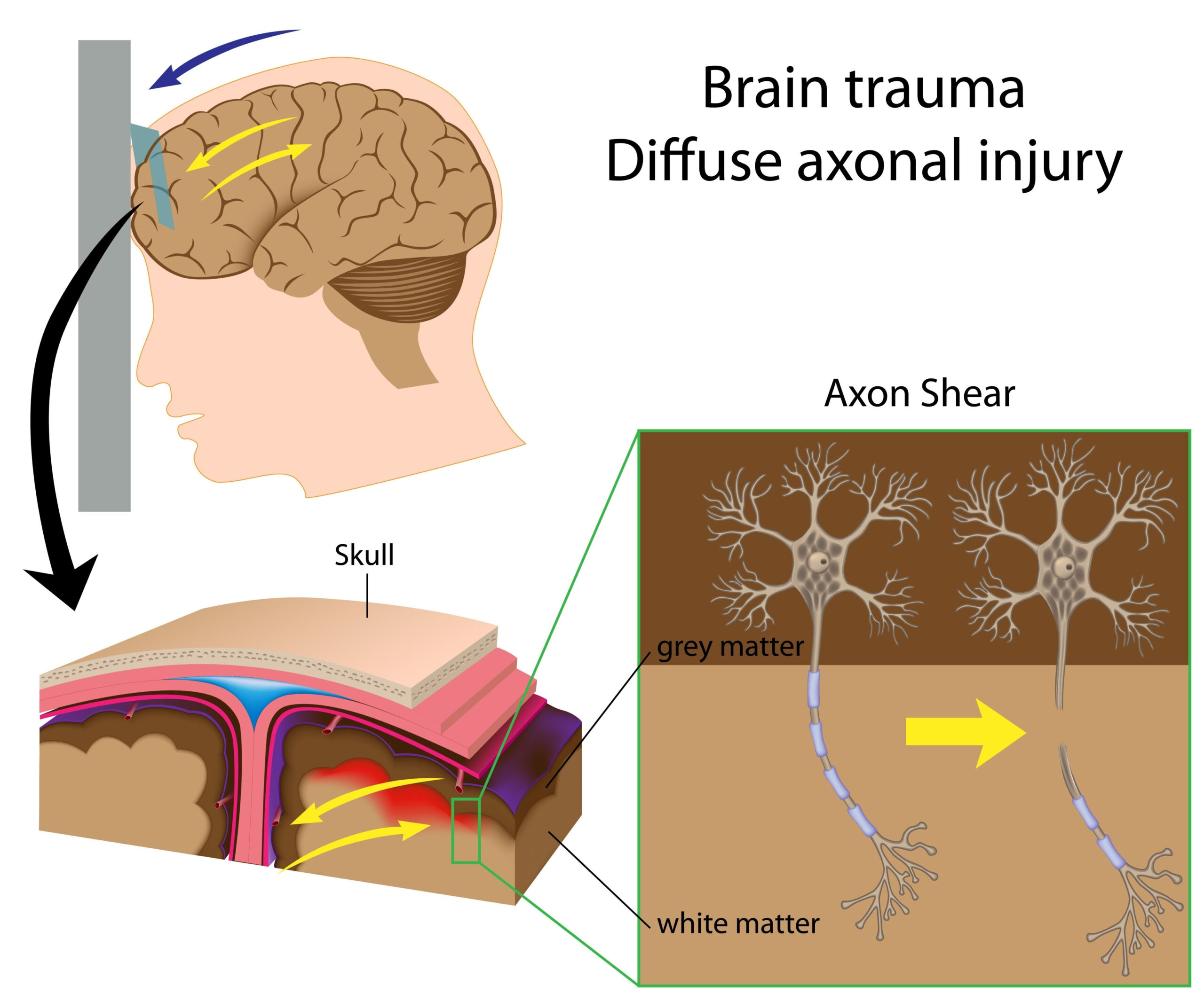
The brain undergoes various structural changes as a result of exposure to blast events, particularly in military personnel such as Special Operations Forces soldiers. These changes are primarily associated with traumatic brain injury (TBI), which has been documented in numerous studies examining the impact of explosive blasts. The unique conditions faced by these soldiers—including high-velocity projectiles, concussive forces, and prolonged exposure to blast waves—can lead to significant neurological consequences.
Research indicates that blast exposure can result in alterations in brain morphology, particularly in regions responsible for cognitive functions, emotional regulation, and motor control. Studies utilizing advanced imaging techniques, such as magnetic resonance imaging (MRI), have identified decreased grey matter volume and alterations in white matter integrity among those experiencing blast injuries. For example, veterans may show differences in the prefrontal cortex, which is crucial for decision-making and emotional responses, and the hippocampus, essential for memory formation.
Additionally, these structural brain changes have been correlated with various psychological conditions commonly seen in blast-exposed soldiers, including post-traumatic stress disorder (PTSD), depression, and anxiety. The interplay between the physiological changes in the brain and the psychological profile of these soldiers raises significant questions about the long-term effects of blast exposure on mental health. Enhanced understanding of the mechanisms underlying these changes is vital for developing targeted interventions and rehabilitation strategies.
Moreover, there is evidence suggesting that the severity of structural brain changes might depend on the magnitude and frequency of blasts experienced. Soldiers exposed to multiple blasts are at increased risk for cumulative brain injury, highlighting the importance of understanding individual histories and exposure levels when evaluating potential neurological outcomes.
The structural changes in the brains of blast-exposed soldiers are complex and multifactorial. Ongoing research is critical to uncover the nuances of these changes and their implications for the health and well-being of affected individuals. Improved diagnostic and therapeutic approaches will depend on further exploration of the relationship between blast exposure and brain health.
Research Design and Methods
The study investigating the brain structural changes in blast-exposed Special Operations Forces soldiers employed a multi-faceted approach to ensure comprehensive data collection and analysis. The research encompassed both qualitative and quantitative methodologies, enabling a holistic view of the impact of blast exposure on neurological health.
Participants included active-duty Special Operations Forces soldiers who had been exposed to explosive blasts during combat or training exercises. Inclusion criteria were set to ensure participants had verified exposure history, as well as access to medical records. A control group comprised soldiers with no history of blast exposure, matched for age, sex, and similar military roles, was established to provide a comparative framework.
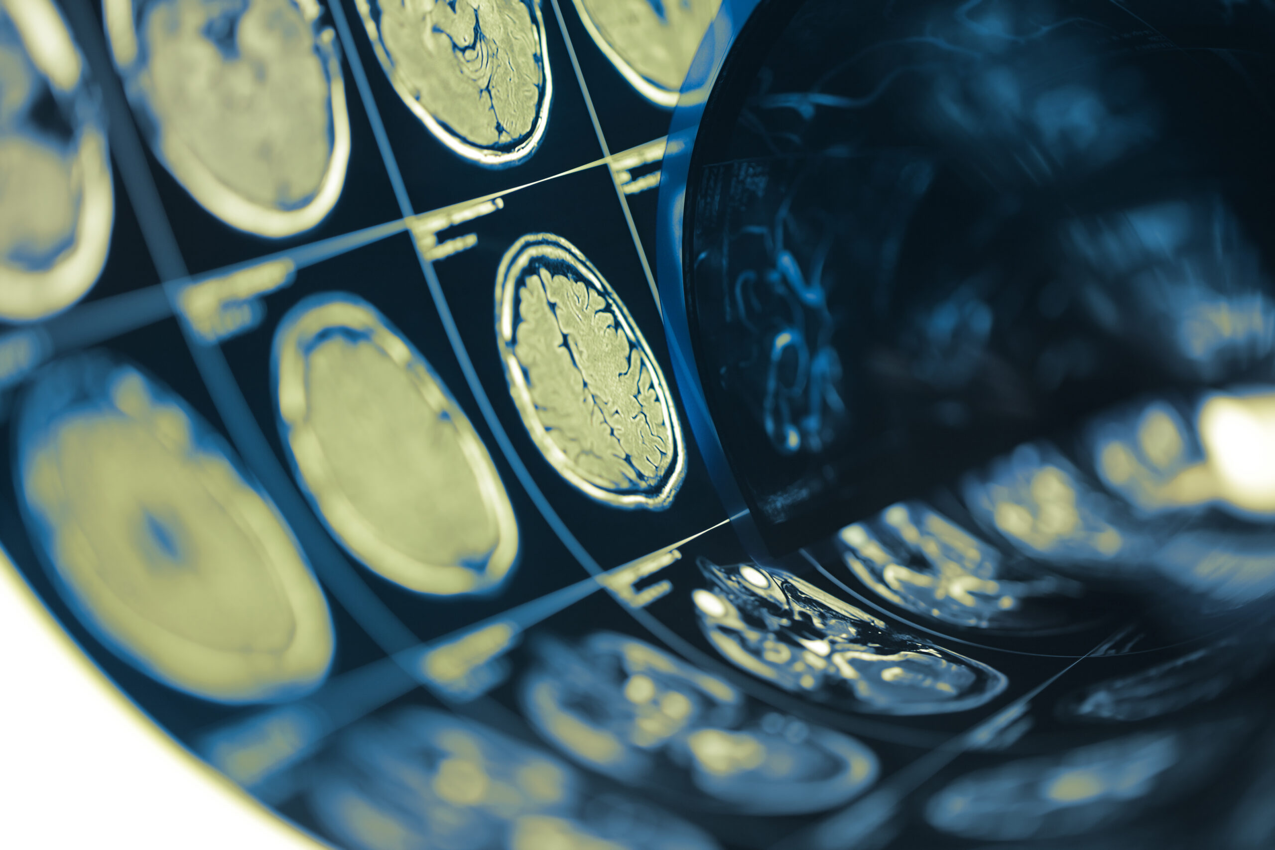
Advanced imaging techniques were employed, notably high-resolution magnetic resonance imaging (MRI), to assess brain structure. The MRI scans were analyzed to identify changes in grey matter volume and white matter integrity. This involved automated morphometric techniques to quantify brain regions, utilizing software like FreeSurfer for accurate volumetric measurements and diffusion tensor imaging (DTI) to evaluate white matter pathways.
To further contextualize the imaging data, neuropsychological assessments were conducted, measuring cognitive functions such as executive function, memory, attention, and emotional regulation. Standard assessments like the Wechsler Adult Intelligence Scale (WAIS) and the Beck Depression Inventory were administered to establish correlations between structural brain changes and psychological conditions such as PTSD, anxiety, and depression.
Additionally, external factors like the history of exposure, including the number of blasts and the intensity of each exposure, were rigorously documented through self-report inventories and military records. This facilitated an analysis of the relationship between exposure patterns and the degree of neurological impact, potentially highlighting a dose-response relationship.
Statistical analyses employed multivariate methods to examine the data, allowing researchers to account for confounding variables such as age, duration of military service, and pre-existing psychological conditions. By using mixed-effects models, the study aimed to discern the specific contributions of blast exposure to the observed brain changes while controlling for individual differences.
Ethical considerations were paramount throughout the study. All participants provided informed consent, and the research was approved by an institutional review board. The privacy and confidentiality of subjects were strictly maintained, ensuring that data handling policies adhered to federal regulations regarding human subjects research.
This rigorous research design sets the stage for not only identifying the extent of brain changes in blast-exposed soldiers but also serves as a framework for future research aimed at understanding the underlying mechanisms of blast-induced neurological injuries. It underscores the importance of integrating diverse research methodologies to capture the complexity of brain health in military personnel.
Results and Analysis
In the analysis of the data collected from this study, significant findings emerged regarding the relationship between blast exposure and structural brain changes among Special Operations Forces soldiers. Participants with documented histories of exposure to explosive blasts exhibited notable reductions in grey matter volume within key brain regions. Specifically, critical areas such as the prefrontal cortex and the hippocampus showed decreased volume compared to the control group, affirming the hypothesis that blast exposure adversely affects brain morphology. The alterations in the prefrontal cortex are particularly concerning given its role in executive function and decision-making, while changes in the hippocampus may underlie memory-related difficulties reported by veterans.
White matter integrity was assessed through diffusion tensor imaging (DTI), revealing significant disruptions in the neural pathways crucial for effective communication between different brain regions. Findings indicated reduced fractional anisotropy in several areas, suggesting that exposure to blasts may contribute to impaired connectivity. This disruption is likely to correlate with cognitive deficits, as effective information processing relies on the robustness of these neural networks.
The neuropsychological assessments further supported the brain imaging results, with many participants reporting difficulties in cognitive functions such as attention and memory. Correlation analyses indicated that the extent of structural brain changes was associated with the severity of psychological conditions, including PTSD and depression. For instance, soldiers with reduced grey matter volume in the prefrontal cortex were more likely to score higher on measures of emotional dysregulation, underscoring the link between physiological brain changes and psychological outcomes.
The study also illuminated the dose-response relationship between the magnitude of blast exposure and brain changes. Participants who experienced multiple blasts showed greater structural alterations compared to those with a single blast exposure. This pattern suggests that repetitive blast events may exacerbate neurological injury, leading to cumulative effects on brain structure and function. Interestingly, the most severe cases of TBI among participants were typically associated with complex exposure histories involving both combat and training environments, emphasizing the importance of a thorough assessment of blast exposure histories in future research.
Statistical models confirmed the significance of these findings, as multivariate analyses accounted for potential confounding variables. Results indicated that even after controlling for age, duration of service, and pre-existing conditions, the impact of blast exposure on brain structure remained significant, reinforcing the idea that these physical changes are indeed attributable to the blasts rather than other factors.
Furthermore, the dimensionality of the changes was reflected in participants’ reports of mental health symptoms. Many soldiers interpreted their cognitive difficulties not just as isolated issues but as part of a broader spectrum of neuropsychological challenges they faced post-exposure. This convergence of structural brain findings with self-reported psychological experiences underscores the complex interactions between brain health and mental well-being.
Drawn together, these results highlight critical implications for the health care of blast-exposed soldiers. Understanding the extent of brain alterations and their associated cognitive and emotional difficulties is essential for guiding effective treatment strategies. The findings call for a multidisciplinary approach to rehabilitation that addresses both the physiological and psychological ramifications of blast exposure, paving the way for enhanced clinical protocols tailored to the unique needs of these military personnel.
Future Directions and Recommendations
Future investigations into brain structural changes resulting from blast exposure in Special Operations Forces soldiers should emphasize longitudinal studies to better understand the trajectory of brain health over time. By following individuals from multiple time points after exposure, researchers can discern how brain alterations evolve and whether they stabilize, progress, or potentially reverse with appropriate interventions.
Additionally, a focus on developing and validating biomarkers for blast-induced neurological injury could enhance diagnostic accuracy. These biomarkers could be derived from advanced neuroimaging techniques or biochemical assays, facilitating earlier detection of neurological changes and allowing for timely interventions. For instance, integrating neuroimaging with blood-based biomarkers might offer a more comprehensive understanding of the physiological impact of blasts on the brain.
Furthermore, exploring the role of genetic and environmental factors in mediating individual susceptibility to blast-related injuries is crucial. Research should investigate which genetic predispositions might influence the severity of brain changes and recovery outcomes. Identifying high-risk groups based on genetic testing could guide personalized treatment plans and preventive strategies.
The clinical implications of these findings highlight the need for enhanced protective measures for soldiers during training and combat operations. Developing equipment that minimizes blast exposure or adjusting training methodologies to limit repetitive exposure could be pivotal in safeguarding brain health. This approach aligns with the concept of primary prevention, aiming to reduce the incidence of TBI before it occurs.
Rehabilitation strategies also require refinement. Cognitive rehabilitative techniques that address specific deficits associated with the identified structural changes in the brain can be developed. Tailoring therapeutic interventions to target areas of cognitive impairment, such as executive function and memory, may lead to better outcomes for affected soldiers. In addition, incorporating psychological support and therapies aimed at addressing co-occurring mental health issues, such as PTSD and depression, is essential for a holistic rehabilitation strategy.
Finally, further research should engage soldiers in the design and implementation of studies. Their insights and experiences can inform more relevant research questions and methodologies, thereby enhancing the applicability and effectiveness of interventions. Community engagement can also foster a supportive environment for veterans, encouraging open discussions about their experiences and the impact of blast exposure on their lives.
Addressing the myriad complexities of blast-induced brain changes in Special Operations Forces soldiers necessitates a multifactorial research approach. By integrating diverse methodologies, focusing on preventive measures, and refining rehabilitation strategies, the healthcare community can better support the neurological health of these service members.