Case Presentation
A 35-year-old male presented to the emergency department after sustaining a traumatic brain injury in a fall from a height of approximately three meters. Upon arrival, he exhibited signs of altered mental status, confusion, and had a Glasgow Coma Scale score of 12. Initial evaluation through a computed tomography (CT) scan of the head revealed a significant left-sided subdural hematoma, prompting urgent neurosurgical consultation.
The patient’s medical history was notable for hemophilia B, a hereditary bleeding disorder caused by a deficiency in clotting factor IX, which profoundly complicates management in trauma cases. Prior to the incident, he had been receiving regular treatment with factor IX infusions, but there was a noted lapse in adherence to his treatment regimen during the two weeks leading up to his injury. This noncompliance raised immediate concerns about his coagulopathy and the potential for excessive bleeding as a result of his condition.
On physical examination, he displayed mild hypertension, with blood pressure readings of 160/95 mmHg, indicating a possible hypertensive emergency, which may have contributed to the injury’s severity. Additionally, further investigation revealed slight swelling over the left parietal region, but no additional injuries were detected upon a full trauma assessment. Laboratory tests showed significantly prolonged activated partial thromboplastin time (aPTT), consistent with his hemophilia diagnosis, while prothrombin time (PT) and platelet function remained within normal limits.
Following initial resuscitation and stabilization, the neurosurgical team opted for a significant intervention due to the size of the hematoma and the neurological compromise evidenced by the imaging results. Preoperative factor IX was administered to correct the coagulopathy before proceeding with a craniotomy for evacuation of the subdural hematoma.
Diagnostic Challenges
The management of patients with hemophilia B presenting with acute neurological insults remains a complex endeavor due to the unique interplay between coagulopathy and trauma-induced complications. In this case, the patient’s underlying bleeding disorder posed significant diagnostic challenges that complicated both clinical assessment and management strategies. Hemophilia B, characterized by a deficiency in clotting factor IX, leads to impaired blood coagulation, thereby increasing the risk of severe bleeding during traumatic events, such as the fall experienced by this patient.
Upon examination, the disparity between the patient’s apparent consciousness level and his prolonged activated partial thromboplastin time (aPTT) raised critical questions about his coagulation status and the potential for substantial intracranial bleeding. The urgency of his situation was compounded by the need to balance immediate surgical intervention for the subdural hematoma with the necessary correction of his coagulopathy. The dilemma was further intensified by the recent lapse in his factor IX infusions; understanding the timeline of this noncompliance was crucial in establishing his current bleeding risk.
The initial CT scan findings indicated significant hematoma, yet the extent of brain injury related to trauma versus that attributable to coagulopathy is often difficult to delineate. Clinicians must weigh the risk of surgical intervention against the potential consequences of delaying treatment in a patient significantly affected by a coagulopathic state. In this case, the decision to proceed with a craniotomy required prompt coordination between the trauma and hematology teams to ensure optimal management of his bleeding disorder prior to surgical intervention.
Additionally, the patient’s mild hypertension, evidenced by elevated blood pressure readings, complicated the clinical picture, potentially signaling elevated intracranial pressure or an underlying chronic condition exacerbated by the acute traumatic event. The interplay between hypertension and intracranial pathology calls for vigilant monitoring and management, as fluctuations in blood pressure can significantly influence bleeding tendencies in patients with hemophilia.
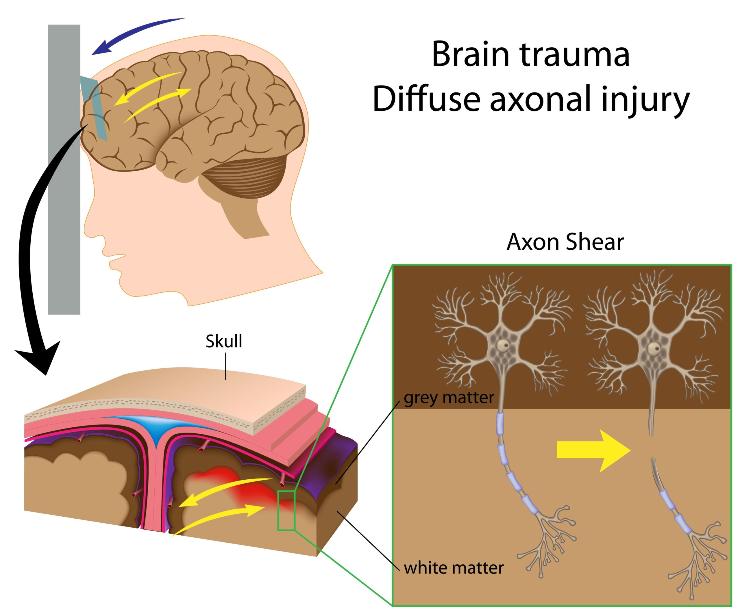
Moreover, the diagnostic process was complicated by the need for additional imaging or assessments. While the CT scan initially provided vital information about the hematoma, further studies might be warranted to evaluate the extent of brain injury and any associated conditions such as contusions or diffuse axonal injury. However, the priority remained the immediate resolution of the life-threatening condition posed by the hematoma, alongside managing the complexities introduced by the patient’s hemophilia B.
The diagnostic pathway during this acute scenario necessitated a multispecialty approach, understanding the intricate balance between addressing traumatic injuries and managing a hereditary bleeding disorder. The integration of hematologic considerations into trauma care is essential to optimize outcomes in similar cases, emphasizing the need for collaboration among surgical and hematology teams in emergency settings.
Treatment Approach
The management of this patient involved a multifaceted treatment strategy tailored to address both the immediate surgical needs and the underlying coagulopathy associated with hemophilia B. Recognizing the urgency of his neurological condition due to the substantial left-sided subdural hematoma, the neurosurgical team acted quickly to stabilize and prepare him for surgery. Preoperative assessment was essential and focused specifically on correcting his coagulopathy to minimize the risk of excessive intraoperative bleeding.
Prior to taking him into the operating room, the decision was made to administer factor IX replacement therapy. This intervention was critical, as it aimed to restore the patient’s clotting ability. The standard protocol for managing patients with hemophilia undergoing surgery involves calculating the required dose of clotting factor based on the patient’s weight and the type of procedure being performed. In this instance, a higher dose was warranted given the significant risk of hemorrhage associated with trauma-related neurosurgery. The patient’s treatment history also necessitated closer monitoring of factor levels to ensure they reached adequate thresholds for safe operative intervention.
Following the administration of factor IX, his coagulation profile was reassessed, and the activated partial thromboplastin time (aPTT) showed significant improvement, indicating that his bleeding risk was mitigated sufficiently for surgery to proceed. Careful monitoring of his hemostatic status is a critical component of treatment in such scenarios, involving repeat laboratory evaluations before and after factor administration.
Once the coagulopathy was addressed, the patient was taken for a craniotomy. The surgery itself involved making an incision and creating a bone flap to access the hematoma. Surgeons meticulously evacuated the subdural hematoma, ensuring that any clotted blood was removed to relieve pressure on the brain. During the procedure, continuous attention was given to hemostasis, with additional factor IX administered as needed to control bleeding risks inherent in the surgical process.
Postoperatively, the patient was closely observed in the intensive care unit (ICU), where he was monitored for neurological status, vital signs, and signs of re-bleeding. The management of his hematological condition continued post-surgery, with additional doses of factor IX given to maintain adequate clotting function during the initial recovery phase. The importance of adhering to a prescribed treatment regimen was reinforced, as ongoing factor administration is crucial in preventing recurrence of bleeding complications.
The multidisciplinary team approach that included trauma surgeons, hematologists, and critical care physicians was vital for optimizing outcomes. The complexity of this case underlined the necessity of collaborative care, particularly for patients with a history of bleeding disorders facing acute medical challenges. Comprehensive follow-up care and patient education regarding hemophilia management post-discharge were also prioritized to reduce the likelihood of future complications and to encourage adherence to treatment protocols.
Conclusion and Recommendations
In addressing the patient’s condition, several recommendations emerge from this case that may help guide future clinical practice for similar presentations involving hemophilia B and traumatic brain injuries. First, it is imperative that emergency departments and trauma centers have protocols in place to quickly identify and manage patients with bleeding disorders. Understanding the unique challenges presented by coagulopathy can facilitate timely decision-making and improve surgical outcomes.
Collaboration among specialists is essential. A robust multidisciplinary approach involving trauma surgeons, hematologists, and anesthesiologists is crucial in managing complex cases like this. Establishing clear communication channels and treatment protocols ahead of time can expedite care and reduce the risk of delays that can jeopardize patient safety.
Specifically, the importance of preoperative factor IX replacement therapy cannot be overstated. Facilities should ensure that they have immediate access to coagulation factor concentrates and that personnel are trained in calculating appropriate dosages based on patient weight and surgical risks. Hematology teams should be integrated into the trauma response team to provide expertise in real-time, particularly for patients with known bleeding disorders.
Education and adherence to treatment in patients with hemophilia are also critical. For this patient, the lapse in factor IX infusions prior to his injury highlighted the need for regular reinforcement of the importance of compliance with treatment regimens. Patient education initiatives should focus on demonstrating the serious consequences of non-adherence and strategies for maintaining consistent treatment, including regular follow-up appointments and potential home administration of factor concentrates.
Additionally, ongoing monitoring of blood pressure in patients with traumatic injuries is vital, particularly in the context of hypertension. Clinicians should be vigilant for signs that may indicate progression of brain injury or secondary complications related to elevated blood pressure. This monitoring will aid in ensuring the timely intervention required to stabilize the patient’s neurological condition post-injury.
Lastly, it is worth considering the implementation of standardized protocols for imaging and assessment of brain injuries in patients with bleeding disorders. Early and thorough evaluation can help delineate between trauma-related damage and complications arising from coagulopathy, allowing for more accurate surgical planning and postoperative care.
This case exemplifies the critical intersections of hematology and trauma surgery while underscoring the necessity for enhanced system preparedness and inter-professional collaboration in clinical practice. By establishing clear protocols and educational frameworks, healthcare teams can significantly improve outcomes for patients facing the dual challenges of acute neurological injuries and bleeding disorders.