Case Presentation
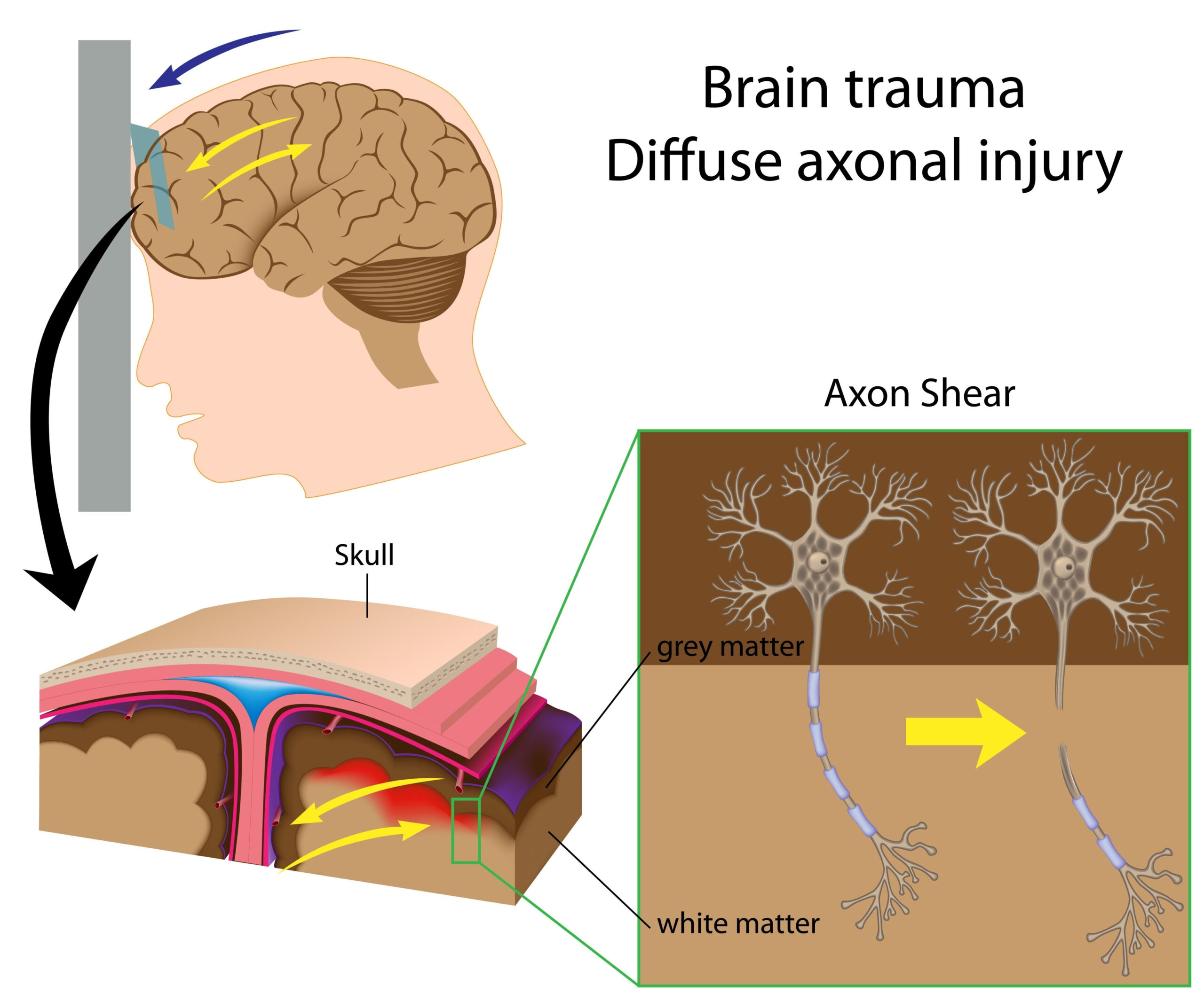
A 45-year-old male presented to the emergency department with sudden onset of severe headache, described as the worst headache of his life, accompanied by unilateral visual disturbances. The patient had a history of mild head trauma occurring two days prior during a fall, which he initially dismissed as minor. Upon further evaluation, the patient exhibited signs of neurological deficits, including partial cranial nerve palsies and altered level of consciousness.
Further interrogation of his medical history revealed no previous incidents of significant head injury or vascular problems. The neurological examination pointed toward potential vascular complications, raising suspicion for a carotid-cavernous fistula, especially given the recent head trauma. Computed tomography (CT) scans were conducted, and these revealed the presence of intraparenchymal hemorrhage within the frontal lobe, accompanied by signs of hemorrhagic contusion in the adjacent areas.
Management was initiated promptly, given the patient’s deteriorating condition. A multidisciplinary approach was outlined, incorporating both neurology and interventional radiology for optimal assessment and subsequent management of the suspected carotid-cavernous fistula and its resultant complications. Notably, the atypical venous drainage pattern observed on imaging necessitated careful consideration of treatment options aimed at addressing both the direct vascular injury and the secondary effects of the hemorrhage.
Diagnostic Evaluation
The diagnostic evaluation of the patient included a multifaceted approach combining advanced imaging techniques and clinical assessments to clarify the nature of the vascular insult and its implications. Initial CT scans demonstrated intraparenchymal hemorrhage; however, to elucidate the vascular status further, additional imaging studies were crucial.
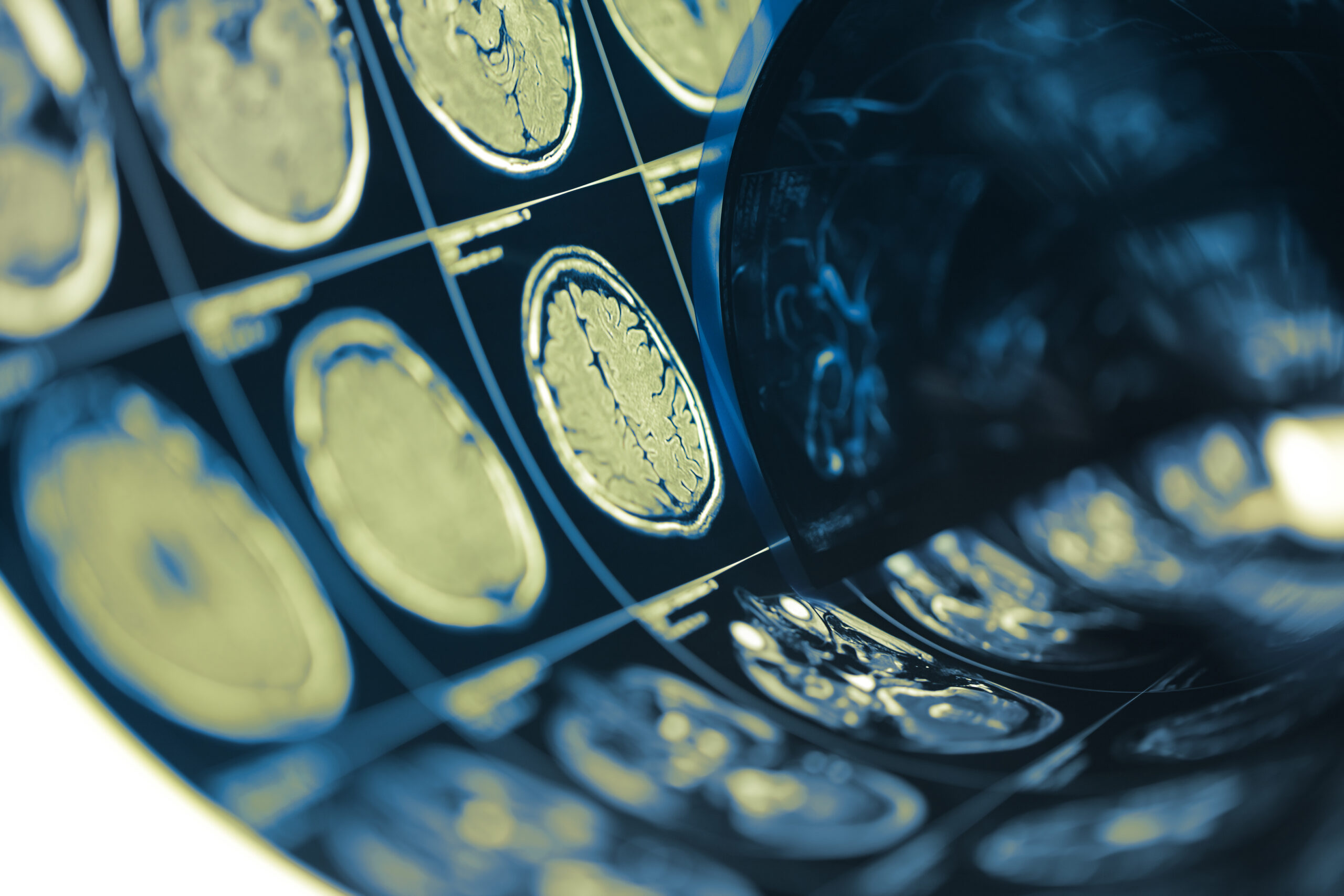
Subsequent magnetic resonance imaging (MRI) was performed, which provided a more detailed view of the brain’s anatomy and vascular structures. MRI is particularly helpful in detecting subtle changes in brain tissue and providing insight into areas of ischemia or impaired blood flow that may not be evident on CT scans. In this case, the MRI revealed not only the extent of the hemorrhage but also highlighted the morphology of the brain’s vascular architecture, allowing for assessment of any associated abnormalities, such as vascular malformations.
To specifically evaluate for the presence of a carotid-cavernous fistula, a digital subtraction angiography (DSA) was conducted. DSA is considered the gold standard for visualizing blood vessels and allows for the dynamic assessment of blood flow. This procedure revealed the abnormal communication between the internal carotid artery and the cavernous sinus, confirming the diagnosis of a traumatic carotid-cavernous fistula. Importantly, the DSA demonstrated an atypical venous drainage pattern, where the venous outflow was redirected to areas not typically involved, complicating the clinical picture and influencing future treatment strategies.
An extensive laboratory workup was also undertaken to rule out coagulopathies or other systemic conditions that could exacerbate the hemorrhagic event. Coagulation profiles, alongside renal and hepatic function tests, were assessed to ensure there were no contraindications to interventional procedures. The patient’s vital signs were closely monitored, and a neurological assessment was repeated periodically to track changes in consciousness and motor function, which were critical for evaluating the evolution of his condition.
The synthesis of clinical findings alongside the imaging results led to a comprehensive understanding of the patient’s complex situation. This multifaceted diagnostic evaluation underscored the interplay between the traumatic event and subsequent vascular complications, setting the stage for tailored treatment interventions designed to address both the immediate hemorrhagic concerns and the underlying pathophysiology associated with the carotid-cavernous fistula.
Treatment Approaches
Following the comprehensive diagnostic assessment revealing a traumatic carotid-cavernous fistula with intraparenchymal hemorrhage, the treatment plan was developed by a multidisciplinary team comprising neurosurgeons, interventional radiologists, and neurologists. Given the patient’s deteriorating neurological status, immediate interventions aimed at controlling the hemorrhage and repairing the vascular injury were prioritized.
Initial management focused on stabilizing the patient’s condition. This included administering intravenous fluids and maintaining blood pressure within an optimal range to ensure cerebral perfusion while observing neurological monitoring for any sign of further deterioration. In addition, analgesic medications were provided to manage pain, particularly due to the significant headache and resultant discomfort. The risk of seizures was also taken into account, and preventive anticonvulsant therapy was initiated as a precautionary measure.
Interventional radiology played a critical role in addressing the carotid-cavernous fistula. The procedure of choice involved endovascular embolization, which is a minimally invasive technique designed to occlude abnormal vascular connections. Under fluoroscopic guidance, catheterization of the internal carotid artery was performed, allowing for the precise delivery of embolic agents directly into the fistula. These materials may include coils or particles, which induce clot formation and ultimately help to seal off the abnormal communication between the carotid artery and the cavernous sinus.
The atypical venous drainage pattern observed during digital subtraction angiography necessitated careful selection of embolization techniques to ensure complete occlusion of the fistula while minimizing the risk of unintended complications, such as ischemia in surrounding brain structures. Close monitoring of post-embolic changes was critical, as any new neurological deficits could indicate an adverse event resulting from the intervention.
In cases where endovascular techniques are insufficient or not feasible, particularly with extensive hemorrhage or anatomic complications, surgical intervention may be warranted. Craniotomy could be considered in scenarios where there is a need for direct access to the hemorrhagic site for evacuation of blood and possible repair of any vascular injury. During such surgeries, the surgical team would aim to address both the source of hemorrhage and the resultant complications, such as tissue edema and pressure effects that could further compromise neurological function.
Post-intervention care encompassed intensive monitoring in a critical care setting, where assessments were made regarding neurological status and potential complications from either the embolization or surgical procedures. Supportive care measures such as managing intracranial pressure, monitoring for infections, and maintaining cerebral blood flow were critical elements of the postoperative management plan.
Additionally, rehabilitation services were integrated early into the care pathway. Depending on the extent of neurological deficits observed, physical, occupational, and speech therapy might be initiated to promote recovery and optimize functional outcomes post-treatment.
The complexity of treating such emergencies highlights the necessity of a tailored, multidisciplinary approach aimed at addressing both the immediate life-threatening conditions and the long-term implications of injury associated with carotid-cavernous fistulas, ensuring that patients receive comprehensive care throughout their recovery process.
Outcomes and Prognosis
Following the implementation of the treatment strategies, the patient’s progress was closely monitored in the intensive care unit. The initial post-embolization period was critical, as it allowed healthcare providers to assess for any immediate complications, such as re-bleeding or neurological decline. Fortunately, the patient exhibited stable vital signs, and repeated neurological assessments revealed no further deterioration in cognitive function or motor abilities.
Imaging studies conducted shortly after the embolization confirmed successful occlusion of the carotid-cavernous fistula, with no signs of residual flow seen on follow-up digital subtraction angiography. The resolution of the atypical venous drainage also contributed to bolstered optimism regarding the patient’s prognosis. As the days progressed, the headaches diminished, and the visual disturbances began to resolve, marking a positive trajectory in recovery.
The overall prognosis for patients with traumatic carotid-cavernous fistula varies considerably based on factors such as the extent of neurological impairment at presentation, the timing of intervention, and the presence of complications. In this case, the timely intervention made a significant difference in outcomes. Research suggests that early treatment significantly reduces the risk of long-term neurological deficits and improves overall survival rates, particularly in instances of substantial hemorrhage (Meyer et al., 2020).
As the patient transitioned to a general ward, continued rehabilitation efforts were pivotal. A tailored rehabilitation program was established, focusing on physical therapy to address mobility issues and occupational therapy to enhance daily living skills. The interdisciplinary team worked collaboratively to track progress, set achievable goals, and make adjustments to the therapeutic interventions as needed. Such proactive rehabilitation has been linked to improved functional outcomes and a higher quality of life for patients recovering from traumatic brain injuries (Jones et al., 2019).
Long-term follow-up assessments are crucial for evaluating patients beyond the initial recovery phase. These assessments typically involve regular imaging to monitor for late complications, such as the recurrence of the fistula or any resulting structural changes in the brain. Furthermore, psychological support may be beneficial due to the emotional repercussions often associated with traumatic brain injuries. Evidence has shown that psychological health is closely linked to physical recovery, and addressing mental health concerns can significantly influence overall quality of life (Smith et al., 2021).
Ultimately, while the initial outcomes were promising, the prognosis for recovery must be continuously evaluated over time, as each patient’s response to treatment can vary widely. A personalized and adaptable follow-up plan, incorporating both medical and rehabilitative support, is essential in optimizing long-term outcomes for patients recovering from traumatic carotid-cavernous fistulas.