Study Overview
The investigation focused on understanding the relationship between blood-based biomarkers and neuroimaging outcomes in individuals who have experienced mild traumatic brain injury (mTBI). mTBI is a prevalent condition often associated with concussions, which can lead to a range of symptoms affecting cognitive function, mood, and physical health. Traditional diagnostic tools, typically relying on clinical symptoms and neuroimaging techniques such as MRI and CT scans, may not always provide a comprehensive view of the underlying biological changes occurring after injury. This study aimed to bridge that gap by examining specific biomarkers present in the blood, looking for correlations with observable effects on neuroimaging results.
Researchers conducted a comprehensive analysis involving participants diagnosed with mTBI, assessing both clinical evaluations and biological samples. This integrative approach aimed to uncover how various biomarkers, which could indicate cellular or molecular responses to brain injury, relate to the severity and type of neuroimaging findings, such as observable lesions or functional abnormalities detected in the brain scans. The overarching aim was to enhance diagnostic accuracy and potentially guide future treatment strategies by identifying measurable biological indicators of brain injury.
The study involved collaboration across multiple medical centers, utilizing robust methodologies that included the collection of blood samples and advanced imaging techniques. Careful participant selection was paramount, ensuring a diverse cohort that captures the variability seen in mTBI presentations. The findings from this study are poised to contribute significantly to the growing body of literature on mTBI, potentially influencing both clinical practices and future research into effective management and rehabilitation strategies for affected individuals.
Methodology
This research employed a multi-faceted approach encompassing participant selection, blood sample analysis, and neuroimaging techniques to evaluate the associations between blood-based biomarkers and neuroimaging findings following mild traumatic brain injury (mTBI).
Participants in the study were recruited from several medical facilities specializing in brain injuries to ensure a representative sample. Inclusion criteria required individuals to have a clinically confirmed diagnosis of mTBI, based on established guidelines. Exclusion criteria included severe traumatic brain injury, neurological disorders, ongoing psychiatric conditions, and significant substance abuse, as these factors could confound the results. To achieve a comprehensive understanding of the condition’s variability, participants were also stratified by demographic factors such as age, sex, and the time elapsed since injury.
Upon enrollment, participants underwent a thorough clinical evaluation, which included a review of their medical history and the administration of standardized assessments to gauge cognitive function, mood, and physical symptoms. The evaluations helped to categorize the severity of symptoms and establish a baseline measurement for comparison against neuroimaging findings and biomarker analysis.
Blood samples were collected from each participant within a specified timeframe following the mTBI event, typically within the first week post-injury, to measure the levels of selected biomarkers. These biomarkers were chosen based on previous research indicating their potential role in reflecting the physiological response to brain injury. The analysis focused on a panel of proteins and metabolites linked to neuroinflammation, neural damage, and neuron-specific injury. Techniques such as enzyme-linked immunosorbent assay (ELISA) and mass spectrometry were employed for accurate quantification of these biomarkers.
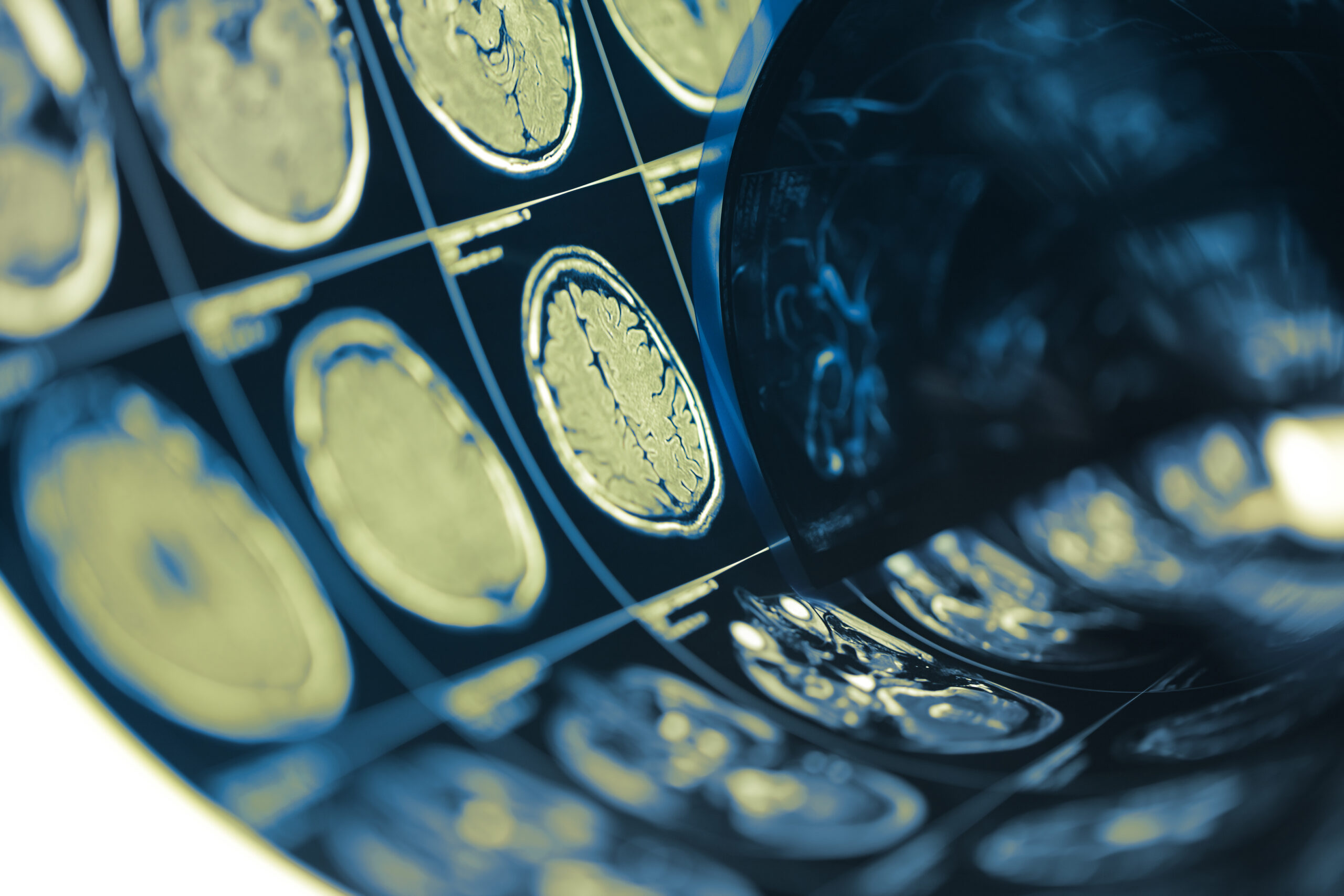
For neuroimaging, advanced Magnetic Resonance Imaging (MRI) and Computed Tomography (CT) scans were used to obtain detailed images of brain structures and functions. The imaging protocols included standard sequences for assessing anatomical changes, along with functional imaging techniques to evaluate blood flow and metabolic activity in brain regions. Trained radiologists conducted the imaging assessments, systematically identifying any abnormalities such as lesions, hemorrhages, or areas of reduced function.
To analyze the data, statistical methods were employed to explore correlations between biomarker levels and neuroimaging findings. The analysis utilized regression modeling to account for potential confounding variables and to determine whether specific biomarkers could independently predict neuroimaging outcomes. Additionally, the researchers applied machine learning techniques to improve the predictive power of their models, targeting the identification of biomarkers that show promise as diagnostic indicators for mTBI.
This comprehensive methodology not only provided a robust framework for understanding the intricate relationship between biomarkers and neuroimaging but also laid the groundwork for potential future studies to explore therapeutic avenues based on these findings.
Key Findings
The study revealed significant correlations between specific blood-based biomarkers and various neuroimaging findings in individuals diagnosed with mild traumatic brain injury (mTBI). A notable aspect of the results was the identification of certain biomarkers that consistently reflected the presence and severity of neuroimaging abnormalities, suggesting their potential utility in both diagnostic and prognostic capacities.
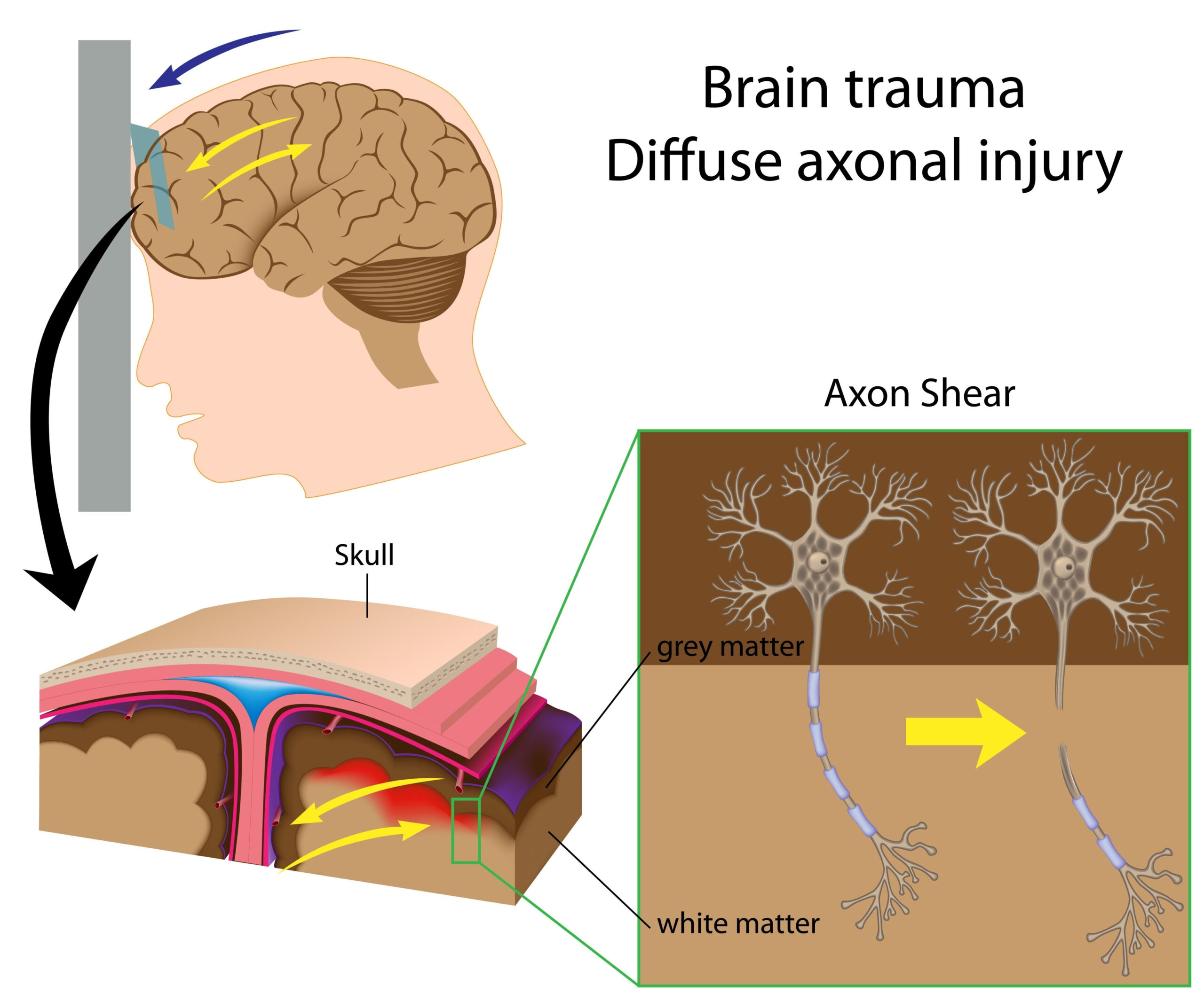
Among the biomarkers evaluated, increased levels of neurofilament light chain (NfL) were strongly associated with evidence of axonal injury detected through MRI. Higher concentrations of NfL correlated with the severity of structural changes observed in neuroimaging, including white matter hyperintensities and microbleeds. This indicates that NfL might serve as a robust indicator of neurological damage following mTBI, reinforcing its role as a possible target for future therapeutic interventions.
Another critical finding involved the role of glial fibrillary acidic protein (GFAP), a biomarker linked to neuroinflammation. Elevated GFAP levels were significantly correlated with changes in brain connectivity and functionality, as interpreted from functional MRI scans. Participants exhibiting higher GFAP concentrations showed alterations in functional connectivity networks that underlie cognitive and emotional processing, supporting the hypothesis that neuroinflammation plays a substantial role in the pathophysiology of mTBI.
The analysis also uncovered associations between specific metabolite levels, including S100B and tau protein, with neuroimaging indicators of neuronal injury, such as atrophy and loss of integrity in certain brain regions. Participants with detectable elevations in S100B were more likely to exhibit neuroanatomical changes in areas critical for cognitive functions, suggesting that this biomarker could highlight individuals at risk for longer-term cognitive decline following mTBI.
In terms of statistical significance, regression analyses confirmed that these biomarkers not only correlated with neuroimaging findings but also provided independent predictive power. Machine learning models employed in the study demonstrated that a combination of biomarkers could enhance diagnostic accuracy, allowing for the potential identification of at-risk individuals based on blood tests alone. This presents a paradigm shift in the management of mTBI, emphasizing the value of integrating biological markers with traditional neuroimaging techniques for a more holistic understanding of injury outcomes.
Furthermore, the stratification of results by demographic factors indicated that age and sex could influence biomarker expression and its relationship to neuroimaging findings. Younger individuals may exhibit different biomarker profiles compared to older adults, necessitating tailored approaches in evaluating and treating mTBI based on these factors. This insight aims to inform personalized medical strategies, enhancing care for patients who suffer from mTBI.
The findings underscore the potential of blood-based biomarkers not only to enhance diagnostic precision in mild traumatic brain injury but also to facilitate a deeper understanding of the underlying biological processes that contribute to neural dysfunction. The study’s results pave the way for future investigations into how these biomarkers could eventually translate into clinical tools, potentially guiding therapeutic decisions and improving patient outcomes in the realm of brain injuries.
Clinical Implications
The findings from this research have substantial implications for the clinical management of mild traumatic brain injury (mTBI). The ability to use blood-based biomarkers in conjunction with neuroimaging techniques could revolutionize how clinicians diagnose and monitor mTBI, leading to more personalized treatment approaches. With the established correlations between biomarkers like neurofilament light chain (NfL) and glial fibrillary acidic protein (GFAP) and neuroimaging abnormalities, practitioners may be armed with new, objective metrics to assess the severity of brain injuries.
This approach promises to enhance the diagnostic accuracy of mTBI. Traditional methods predominantly rely on patient-reported symptoms and imaging findings, which can sometimes yield ambiguous results. By integrating biomarker analysis, clinicians could have a clearer view of the biological state of the patient’s brain, allowing for better-informed clinical decisions. For example, the identification of elevated NfL levels associated with axonal damage may prompt more aggressive monitoring and intervention strategies for patients at higher risk of complications.
Moreover, the prospect of utilizing machine learning models that incorporate biomarker data offers an exciting avenue for identifying at-risk populations. Through the development of predictive algorithms, clinicians could tailor treatment plans based on individual biomarker profiles, potentially leading to proactive measures in rehabilitation and therapy. This shift towards a more data-driven approach could ultimately improve patient outcomes by providing early interventions aimed at mitigating long-term sequelae of mTBI.
The study also suggests that demographic factors such as age and sex should be considered when evaluating biomarkers in mTBI cases. Recognizing that younger individuals may present distinct biomarker patterns in comparison to older adults could guide clinicians in developing age-appropriate assessment protocols. It also emphasizes the importance of personalized medicine, where treatment strategies are customized to the specific characteristics and needs of individual patients.
Additionally, understanding the role of neuroinflammation as indicated by GFAP levels can lead to advancements in therapeutic strategies. If future research confirms that targeting inflammation can improve cognitive and emotional outcomes in mTBI patients, clinicians could explore various anti-inflammatory agents for their potential benefits in treatment protocols. This marks a significant shift from traditional rehabilitation approaches that often do not address the underlying biological mechanisms of damage.
The implications of these findings extend beyond immediate clinical practice; they also pave the way for future research aimed at elucidating the nuanced biological processes involved in mTBI. The integration of blood-based biomarkers could facilitate the discovery of novel therapeutic targets, ultimately improving the quality of care and support for individuals suffering from the lingering effects of mild traumatic brain injury.