Study Overview
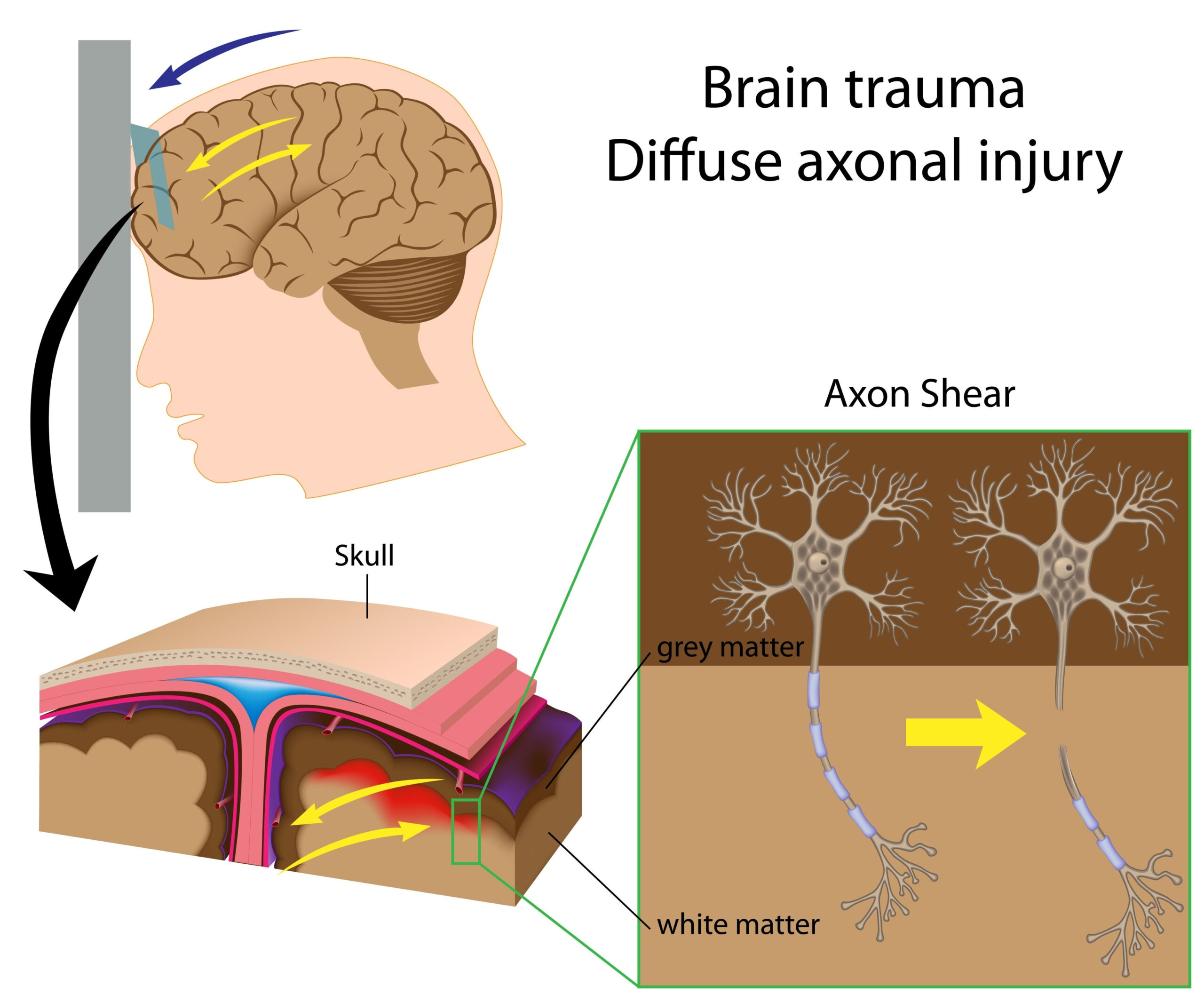
The research focused on the alterations in water channel dynamics in astrocytes following the administration of buprenorphine in a rat model that simulates diffuse traumatic brain injury (TBI). As traumatic brain injuries often lead to significant neurological deficits, understanding the cellular and molecular mechanisms behind these changes is vital for developing effective therapeutic approaches. Astrocytes, a type of glial cell in the brain, play a crucial role in maintaining homeostasis, supporting neuronal function, and regulating water balance within the central nervous system. The study specifically investigated the response of astrocytic water channels, particularly aquaporin-4 (AQP4), in the context of buprenorphine treatment amid the complications that arise from TBI.
Buprenorphine is an opioid analgesic known for its agonistic effects on mu-opioid receptors and antagonistic properties on kappa-opioid receptors. The dual nature of buprenorphine presents a unique angle to explore its neuroprotective potential, particularly in injuries where inflammation and edema are prevalent. Given the involvement of astrocytic water channels in regulating edema post-injury, it was hypothesized that buprenorphine might modulate AQP4 expression and localization, thereby influencing water transport and cellular responses after TBI.
The study utilized a well-defined experimental protocol with controlled administration of buprenorphine and comprehensive analysis of tissue samples. This approach aimed to elucidate the interaction between astrocytes and water channels in the aftermath of brain injury and how pharmacological intervention could modify these interactions, potentially paving the way for novel therapies in TBI management.
Methodology
The research employed a rat model of diffuse traumatic brain injury to investigate the specific changes in astrocyte-associated water channels following buprenorphine administration. This approach allowed for the simulation of TBI in a controlled laboratory setting, providing a framework to analyze how pharmacological treatment could alter cellular responses to injury.
The study began with the induction of diffuse TBI using a standardized impactor device, which creates consistent and reproducible brain injury. Following the injury, the rats were randomly assigned to receive either buprenorphine or a saline control treatment. Buprenorphine was administered in clinically relevant doses, reflecting concentrations used in human medical scenarios, to appropriately assess its potential neuroprotective effects. Control animals received saline injections to account for the effects of handling and injection procedures.
To assess the effectiveness of buprenorphine treatment, the research team conducted a series of behavioral and physiological assessments. These included tests for motor coordination and cognitive function, which provided insight into the overall impact of the injury and the therapeutic intervention. Behavioral assessments were performed at multiple time points post-injury to capture the progression of recovery and the potential benefits of buprenorphine administration.
Following behavioral testing, the researchers collected brain tissue samples for detailed analysis of astrocytic water channels, focusing particularly on aquaporin-4 (AQP4). The tissue samples underwent histological analysis and immunohistochemistry to evaluate the localization and expression levels of AQP4 across different treatment groups. This involved using specific antibodies that bind to AQP4, allowing for visualization under a microscope.
Moreover, quantitative analysis of AQP4 expression was conducted through western blotting and real-time polymerase chain reaction (PCR), providing complementary data on protein and gene expression changes induced by buprenorphine. These methodologies enabled the researchers to ascertain both the spatial distribution and the overall abundance of AQP4 in the context of brain injury and buprenorphine treatment.
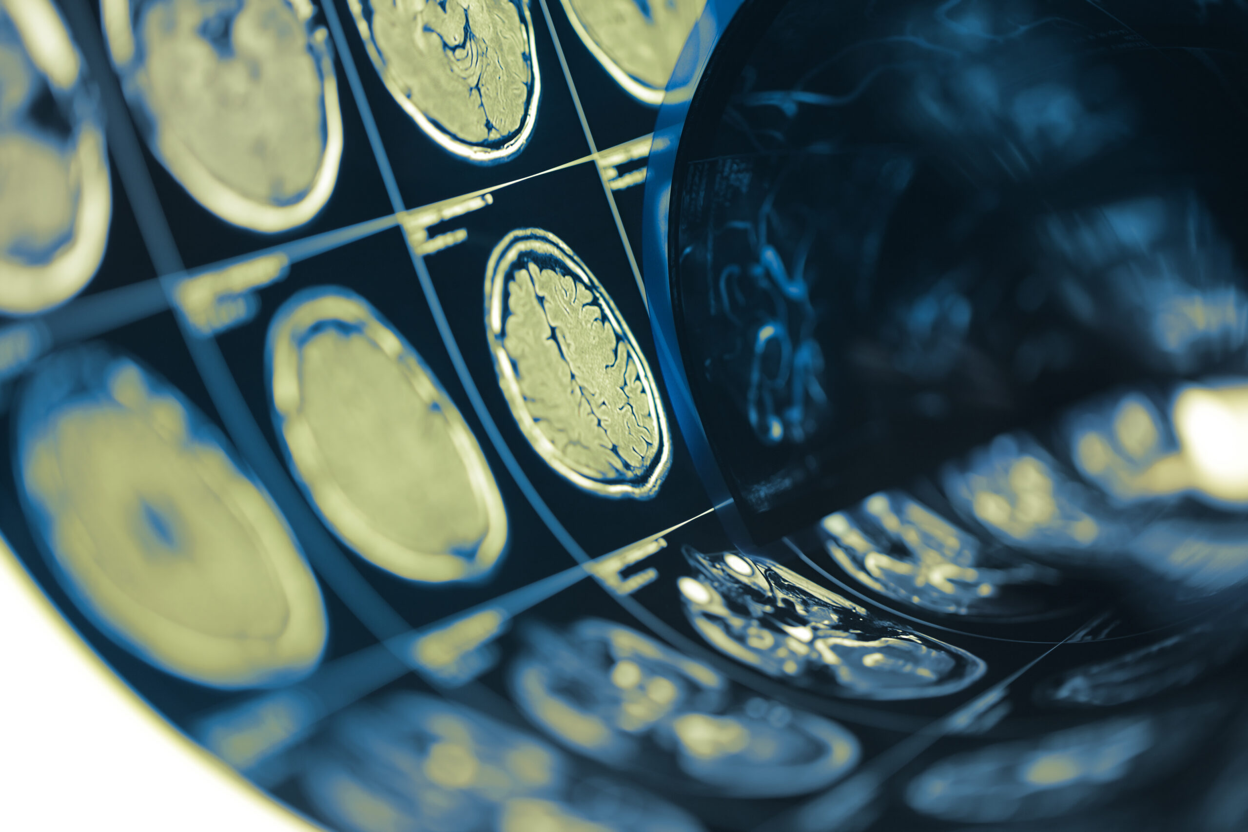
In addition to molecular assessments, tissue edema was quantitatively assessed using MRI imaging techniques, which provided insights into changes in water content within the brain tissue post-injury. This imaging modality allowed for non-invasive observation and quantification of edema, supporting the investigation into how buprenorphine treatment modulated fluid dynamics in the injured brain.
Through this multifaceted methodology, the study aimed to provide a comprehensive understanding of how buprenorphine influences astrocyte function and water channel dynamics in the complex environment of diffuse traumatic brain injury, ultimately seeking to identify potential therapeutic avenues for improving patient outcomes in TBI.
Key Findings
The study revealed significant alterations in the expression and localization of aquaporin-4 (AQP4) in astrocytes following buprenorphine treatment in a rat model of diffuse traumatic brain injury (TBI). Notably, the administration of buprenorphine led to an upregulation of AQP4 protein levels when compared to the saline control group. Immunohistochemical analyses showed a marked increase in AQP4 staining intensity in areas adjacent to the injury site, suggesting enhanced water channel expression in response to treatment. This finding aligns with the hypothesis that buprenorphine might promote astrocyte activity in mitigating edema following TBI.
Quantitative analyses, including western blotting and real-time PCR, confirmed the increase in AQP4 expression levels. These methods indicated a statistically significant elevation in both protein and mRNA levels of AQP4, reinforcing the notion that buprenorphine not only influences the protein synthesis of water channels but may also enhance gene expression associated with their function. This dual mechanism highlights buprenorphine’s potential as a modulator of fluid transport in the injured brain.
MRI imaging conducted during the study provided essential insights into the effects of buprenorphine on tissue edema. The non-invasive nature of MRI allowed for real-time monitoring of water accumulation within the brain. Following buprenorphine treatment, the measurements indicated a significant reduction in edema volume compared to the controls, suggesting that AQP4 modulation may play a crucial role in facilitating water clearance from the brain tissue. The relationship between reduced edema and elevated AQP4 expression underscores the importance of astrocytic water channels in managing fluid balance during the acute phase of brain injury.
Furthermore, behavioral assessments demonstrated improved motor coordination and cognitive performance in the buprenorphine-treated group, correlating with the biochemical findings. These observations suggest that the neuroprotective effects of buprenorphine are not solely attributable to changes in astrocyte water channel dynamics but may also involve complex neurochemical interactions that support functional recovery following TBI.
Overall, the findings indicate that buprenorphine significantly influences the expression and function of key astrocytic water channels, providing a potential therapeutic pathway for managing post-injury edema and enhancing functional recovery in patients with traumatic brain injuries. The evidence supports further exploration of buprenorphine not only as an analgesic but as a candidate in neuroprotection and treatment for TBI-related complications.
Clinical Implications
The implications of this study extend into several critical aspects of clinical practice regarding the treatment of traumatic brain injury (TBI). The clear upregulation of aquaporin-4 (AQP4) expression following buprenorphine administration suggests that targeted pharmacological interventions may offer a dual benefit: managing pain while also promoting better neurological recovery. This could be particularly significant in acute TBI settings where both analgesia and neuroprotection are vital for improving patient outcomes.
Given the invasive nature of some pain management strategies, buprenorphine’s potential to reduce edema, as evidenced by the reduced volume of tissue swelling seen in MRI assessments, could support the development of less invasive and safer treatment protocols for managing TBI. Efficient water clearance from the brain may mitigate secondary injury due to edema, potentially decreasing the risk of lasting neurological deficits. This is especially relevant for the treatment of patients at high risk for complications stemming from increased intracranial pressure.
Moreover, enhancing AQP4 function could fine-tune the inflammatory response following TBI, linking the physiological response to a more favorable recovery trajectory. The relationship between astrocytic water channel activity and neuronal health underscores the need for further research into the timing and dosage of buprenorphine administration post-injury. Clinicians may find it beneficial to integrate such treatment protocols into acute care guidelines, especially as we better understand the optimal conditions under which buprenorphine exerts its protective effects.
In light of observations regarding improved cognitive and motor function in buprenorphine-treated rats, similar benefits may translate to human patients experiencing TBI. Effective recovery from cognitive impairment, frequently observed post-injury, is a primary focus of rehabilitation efforts. Therefore, integrating buprenorphine into treatment regimens could not only alleviate acute pain but also support complex neuronal recovery processes, enhancing overall rehabilitation outcomes.
Additionally, this study may inform prescriptive practices regarding opioid use in clinical settings. As buprenorphine possesses a favorable safety profile compared to traditional opioids, it may present an attractive option for managing pain in TBI patients, who are often vulnerable to the adverse side effects associated with opioid therapy, including dependency and respiratory distress. Its unique pharmacological properties position it as an effective alternative that could help mitigate these risks while simultaneously providing therapeutic benefits to brain health.
Overall, the research indicates a promising avenue for the use of buprenorphine not merely as an analgesic agent but as a potential neuroprotective treatment that may enhance recovery from traumatic injuries in the brain. Future clinical trials are warranted to further delineate the therapeutic window, optimal dosages, and long-term outcomes associated with buprenorphine use in patients following TBI, guiding effective clinical practices in this challenging domain of medicine.