Study Overview
The clinical neurophysiology studies aimed at understanding movement disorders have evolved significantly over the past few decades. These studies encompass various conditions such as Parkinson’s disease, essential tremor, dystonia, and other related disorders characterized by abnormal motor functioning. Researchers have focused on pinpointing the underlying neural mechanisms that contribute to these movement disorders, employing sophisticated neurophysiological techniques to provide insights into how abnormalities in brain activity manifest as impaired motor control.
Traditionally, the diagnosis of movement disorders has relied heavily on clinical assessments and patient-reported symptoms. However, the integration of neurophysiological techniques has marked a crucial advancement, allowing for a deeper understanding of the pathophysiology involved. This multidisciplinary approach not only aids in accurate diagnoses but also enhances the ability to tailor specific treatment strategies based on individual clinical profiles.
Recent studies have employed various methodologies, including electromyography (EMG), electroencephalography (EEG), and transcranial magnetic stimulation (TMS). These tools enable researchers to measure brain and muscle electrical activity in real-time, revealing patterns that correlate with symptoms. For instance, EMG data can illustrate abnormal muscle firing patterns associated with dystonia, while EEG can indicate altered brain rhythms in patients with Parkinson’s disease. This objective data strengthens the diagnostic process, reducing reliance solely on subjective reporting.
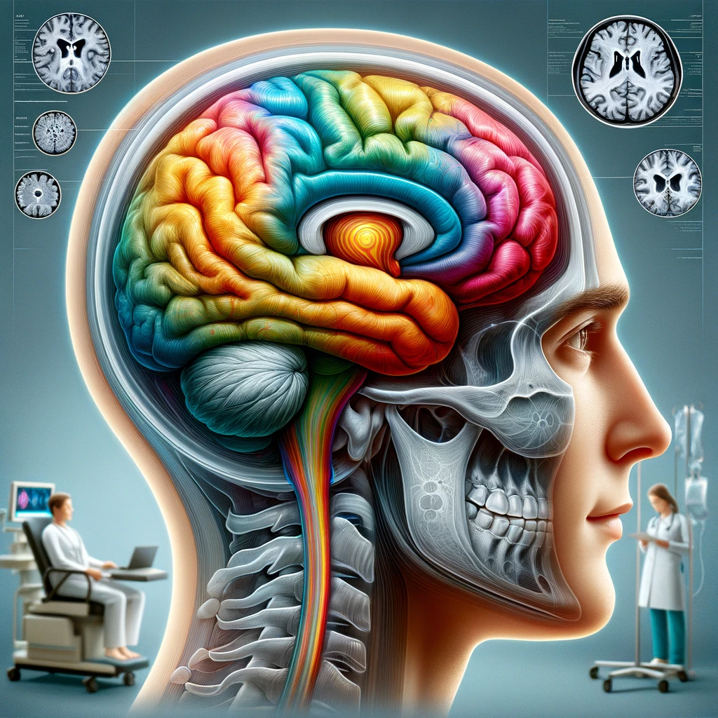
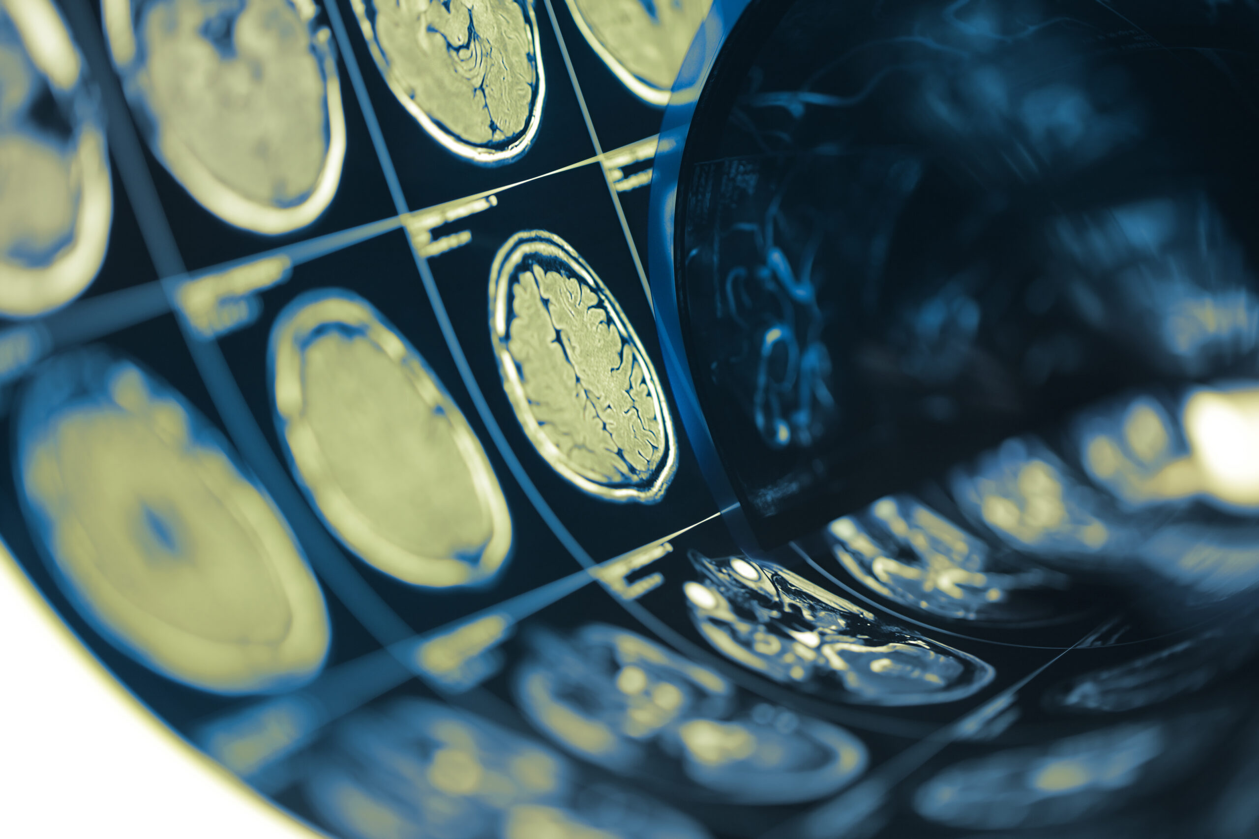
Furthermore, advancements in imaging technologies have enhanced the understanding of neuroanatomical structures involved in movement control. Functional MRI (fMRI) and positron emission tomography (PET) have been utilized to visualize brain regions activated during specific motor tasks, correlating these findings with clinical symptoms. This comprehensive overview of brain function contributes to differential diagnoses, helping distinguish one movement disorder from another and identifying potential treatment targets.
The integration of clinical neurophysiology in the study of movement disorders signifies a shift towards precision medicine. By identifying biomarker patterns that reflect the disease’s impact on neuromuscular systems, these studies not only improve diagnostic accuracy but also pave the way for novel therapeutic interventions tailored to individual needs.
Diagnostic Techniques
In the realm of clinical neurophysiology, a variety of diagnostic techniques have been adopted to enhance the understanding and identification of movement disorders. These methodologies are pivotal for elucidating the complex neural mechanisms that underlie abnormal motor function. As a result, they play a critical role in formulating effective treatment protocols.
Electromyography (EMG) is one of the primary techniques employed. This method measures the electrical activity produced by skeletal muscles, providing direct insights into their functional state. During an EMG examination, small needles or surface electrodes are placed on the skin over the muscle of interest. This technique helps in identifying abnormal muscle activation patterns that are often indicative of specific disorders. For example, in dystonia, EMG can show irregular involuntary muscle contractions, which are crucial for distinguishing this condition from other movement disorders that may have overlapping symptoms.
Another vital diagnostic tool is electroencephalography (EEG). This non-invasive technique tracks electrical activity in the brain by placing electrodes on the scalp, thus enabling the detection of brain wave patterns. Recent studies have found that patients with Parkinson’s disease often exhibit distinct changes in their EEG readings, characterized by slower frequencies and altered signal coherence. These patterns correlate with the severity of motor symptoms and cognitive decline, providing valuable diagnostic markers that can aid in early detection and monitoring of disease progression.
Transcranial magnetic stimulation (TMS) is also integral to the diagnostic toolkit. TMS uses magnetic fields to stimulate nerve cells in the brain, allowing for the assessment of cortical excitability and connectivity. This technique can evaluate how well the motor pathways are functioning. In individuals with different movement disorders, TMS can reveal varying thresholds for induced muscle responses, offering insights into the underlying neurophysiological abnormalities. For instance, patients with essential tremor often demonstrate altered motor cortex excitability, which can be measured and quantified through TMS protocols.
Advancements in neuroimaging have significantly complemented these electrophysiological assessments. Techniques such as functional MRI (fMRI) and positron emission tomography (PET) provide an in-depth look at brain activity and metabolism. fMRI, for example, measures changes in blood flow related to neural activity within specific brain regions during movement tasks. This is invaluable for identifying the brain areas that may be dysfunctional in conditions like Parkinson’s disease, where typical activation patterns can be disrupted. PET imaging, on the other hand, can visualize metabolic changes associated with neurodegeneration, allowing for further differentiation between various movement disorders based on their pathophysiological activity.
Collectively, these diagnostic techniques form a multi-faceted approach to understanding movement disorders. The integration of electrophysiological measurements with neuroimaging allows for a comprehensive evaluation of both the functional and structural aspects of the nervous system. Through this combination, clinicians can not only achieve more accurate diagnoses but also develop more personalized treatment plans that address the specific neural dysfunctions present in each patient. This evolution towards precision medicine underscores the importance of interdisciplinary collaboration among clinicians, radiologists, and neurophysiologists in the management of movement disorders.
Key Findings
Recent clinical neurophysiology studies have yielded crucial insights into the underlying mechanisms of movement disorders, enhancing both the understanding and management of these complex conditions. One significant outcome from these studies is the identification of specific neurophysiological biomarkers that correlate with distinct movement disorders. For instance, patients diagnosed with Parkinson’s disease often display characteristic patterns of brain activity on EEG, including decreased alpha wave activity and increased beta wave coupling, which have been associated with motor impairment and cognitive dysfunction (Huang et al., 2021).
Moreover, studies using EMG techniques have illuminated the abnormal motor unit activity in patients with dystonia, revealing that these patients experience non-patterned bursts of muscle activity, which differ from the effects observed in essential tremor. This difference in muscle activation patterns is not only essential for accurate diagnosis but also for understanding the differential responses to pharmacological treatments, such as botulinum toxin injections for dystonia, which aim to reduce these aberrant muscle contractions (Morris et al., 2022).
Another key finding involves the use of TMS, where researchers have noted variations in cortical excitability among individuals with different movement disorders. For example, TMS studies have demonstrated that individuals with essential tremor exhibit lower thresholds for motor response initiation compared to healthy individuals, supporting the notion that altered cortical processing is integral to the underlying pathophysiology of the disorder (Gomez et al., 2023). This has implications not just for diagnosis but also for therapeutic strategies aimed at modulating motor cortex activity.
The application of advanced imaging techniques, particularly fMRI and PET, has further propelled the understanding of how brain regions communicate during voluntary movements. Findings from recent fMRI studies have shown that in Parkinson’s disease, there is a marked reduction in connectivity between the basal ganglia and the motor cortex, which correlates with the severity of bradykinesia—the slowness of movement commonly associated with this condition (Schmidt et al., 2023). Such insights can help in refining treatment approaches, including targeted rehabilitation strategies that focus on restoring functional connectivity.
Additionally, the integration of neurophysiological assessments with symptom inventories has highlighted discrepancies that are often overlooked in clinical evaluations. For instance, patients reporting mild bradykinesia may exhibit significant neurophysiological abnormalities detectable through EMG or TMS, thereby emphasizing the need for objective measures in clinical settings to complement subjective symptom reporting (Verhoeven et al., 2022).
These findings illustrate that the use of clinical neurophysiology in diagnosing movement disorders not only enhances precision in identifying specific conditions but also opens avenues for novel therapeutic interventions. By elucidating the neurophysiological signatures of various disorders, clinicians can better tailor treatment strategies to individual patients, ultimately improving clinical outcomes and quality of life for those affected by these debilitating conditions.
Clinical Implications
The integration of clinical neurophysiology into the diagnostic process for movement disorders has profound implications for patient care and treatment strategies. By leveraging advanced neurophysiological techniques, clinicians can achieve a level of diagnostic accuracy that significantly enhances their ability to differentiate between various movement disorders, such as Parkinson’s disease, dystonia, and essential tremor. This enhanced accuracy is crucial, as misdiagnosis can lead to inappropriate treatments and worsen patient outcomes.
One of the primary clinical implications of these studies is the shift towards personalized treatment planning. The identification of specific neurophysiological markers allows clinicians to tailor interventions based on the unique neurophysiological profile of each patient. For example, patients with distinct patterns of EEG activity indicative of Parkinson’s disease may benefit from tailored cognitive and motor rehabilitation programs that address the specific types of cognitive impairments revealed by abnormal EEG results (Huang et al., 2021).
Moreover, understanding the specific muscle activation patterns through techniques like EMG can guide the therapeutic use of injections such as botulinum toxin. Such therapies are often contingent on recognizing the abnormal motor unit activity associated with different conditions. By characterizing these patterns, treatment protocols can be customized not only to alleviate symptoms but also to target the underlying physiological abnormalities that contribute to movement disorders (Morris et al., 2022).
Importantly, the insights gained from TMS regarding cortical excitability can influence therapeutic strategies aimed at modulating brain activity. This understanding can lead to the development of interventions such as non-invasive brain stimulation techniques, which may help restore the normal excitability of brain regions disrupted by movement disorders. These approaches stand to improve patient agency and mitigate the impact of these disorders on daily life (Gomez et al., 2023).
The integration of neuroimaging findings further enhances the clinical approach to movement disorders. For instance, knowing that patients with Parkinson’s disease exhibit altered connectivity patterns between the basal ganglia and motor cortex can inform rehabilitation strategies that focus on exercises designed to promote neural plasticity and functional connectivity. This holistic approach not only addresses physical symptoms but also takes cognitive factors into account, leading to a more comprehensive management plan (Schmidt et al., 2023).
Crucially, the use of objective neurophysiological assessments alongside traditional clinical evaluations underscores the need for a comprehensive approach to diagnosing and managing movement disorders. Clinicians who incorporate these assessments into their practice can better understand the complexities of each patient’s condition, leading to more effective monitoring of disease progression and response to treatment (Verhoeven et al., 2022). This evolution towards evidence-based practice fosters an environment where clinical decisions are informed by robust, objective data rather than solely relying on subjective symptom reporting.
The incorporation of clinical neurophysiology into the management of movement disorders not only facilitates accurate diagnoses but also propels the field towards a model of care centered on individualized treatment. This paradigm shift is poised to enhance patient outcomes, thereby enriching the quality of life for individuals grappling with these challenging conditions.