Neurophysiologic Techniques Overview
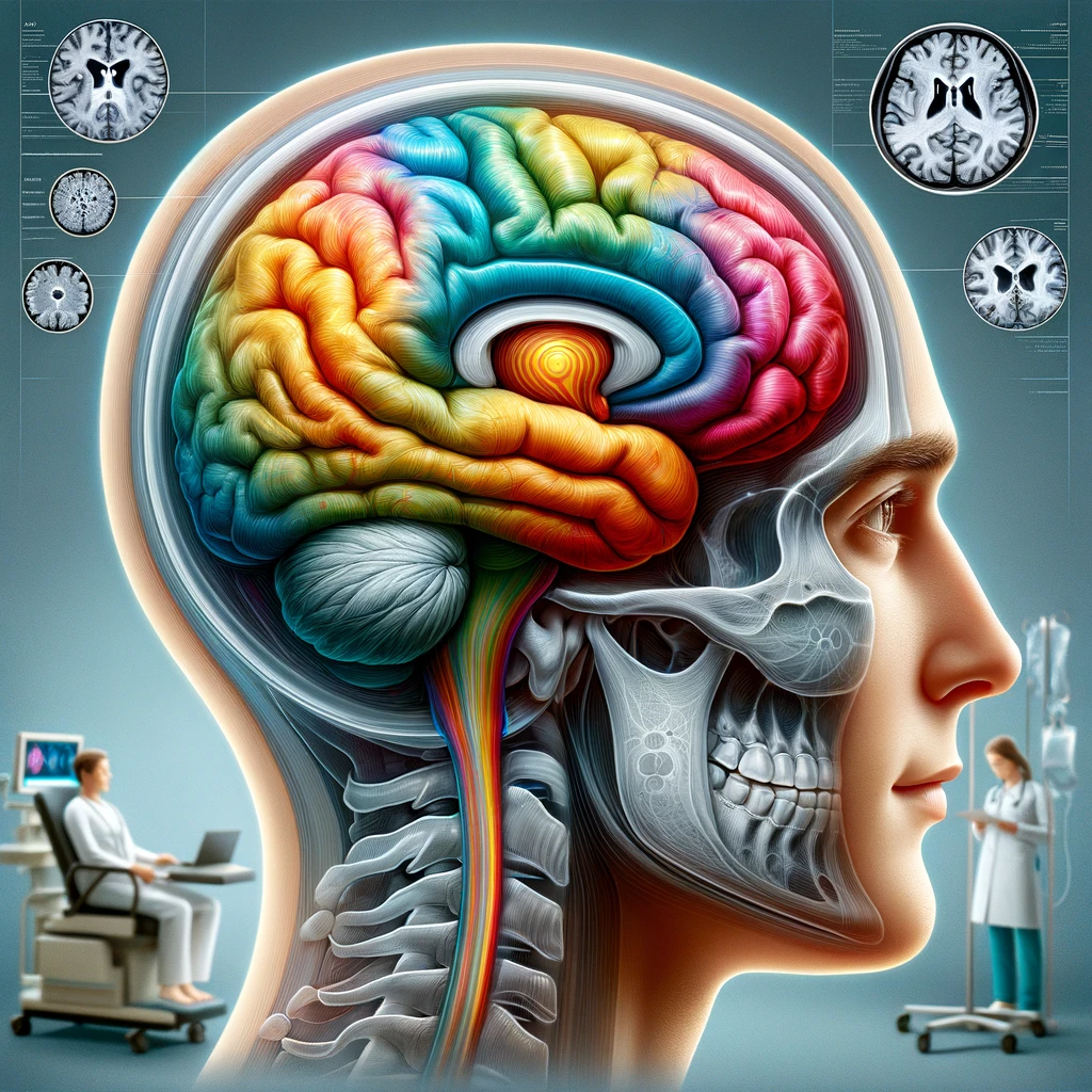
Neurophysiologic techniques are crucial for understanding the complexities of movement disorders, providing insights into the electrical activity of the nervous system during various motor tasks. These methods focus on capturing and analyzing electrical signals generated by neurons, which are pivotal in coordinating bodily movements. They offer the ability to study both the central nervous system and peripheral motor pathways, thereby revealing the intricate interplay that occurs in motor control.
At the heart of neurophysiologic research are techniques that range from surface electromyography (sEMG) to advanced signal processing methodologies. These techniques allow researchers to examine muscle activity and the timing of neuronal firing, which are essential for assessing functional impairments in patients with movement disorders. For instance, measuring electrical signals from muscles can help identify abnormal neuromuscular function, which may be linked to conditions such as Parkinson’s disease, dystonia, or essential tremor.
One of the key aspects of these techniques is their ability to capture temporal changes in neural activity. This dynamic evaluation is critical as movement disorders often involve fluctuating symptoms that can vary greatly over time. With the aid of sophisticated software and analytical methods, researchers can create detailed time-series data that reflect the precise moments of muscle activation, providing a clearer picture of the underlying pathophysiology.
Additionally, neurophysiologic techniques facilitate the exploration of brain activity in relation to movement. For example, coupling surface electromyography with functional imaging modalities can provide a multi-dimensional approach to understanding how brain regions communicate and coordinate motor functions. This integrated perspective helps in identifying specific alterations in motor control, making it a valuable resource for both diagnosis and treatment planning.
The adaptability of these techniques also allows for their application in various contexts, from clinical settings to experimental laboratories. This flexibility enhances their utility in studying both acute and chronic movement disorders, ultimately contributing to the refinement of therapeutic strategies aimed at alleviating debilitating motor symptoms.
In summary, the diverse array of neurophysiologic techniques offers a robust platform for investigating the complexities of movement disorders. By capturing and analyzing neural and muscular activity, these methods provide valuable insights into the mechanisms of movement control and the functional impairments associated with various neurological conditions.
Polymyography Applications
Polymyography, encompassing both surface and intramuscular electromyography, serves as a potent tool in the evaluation of motor function in various movement disorders. Unlike standard electromyography, which primarily focuses on individual muscle action potentials, polymyography captures and analyzes the coordinated electrical activities of multiple muscles simultaneously. This approach allows for a more comprehensive understanding of muscle recruitment patterns and their relationship to movement disorders.
One of the significant applications of polymyography is in the assessment of dystonic movements. Dystonia, characterized by sustained muscle contractions leading to abnormal postures and movements, can be intricately analyzed through polymyographic recordings. Researchers can observe how different muscle groups interact during sustained muscle contractions or during voluntary movements, helping to delineate abnormal activity patterns. By comparing the muscle activation sequences between healthy individuals and those with dystonia, clinicians can identify specific neural pathways that may be disrupted in dystonic patients. This understanding can guide targeted treatments such as botulinum toxin injections or physical therapy tailored to improve motor function.
In cases of Parkinson’s disease, polymyography proves invaluable in examining the bradykinesia, or slowness of movement, that many patients experience. Through detailed recordings of muscle activity during tasks that require precise motor control—such as grasping or walking—researchers can quantify the extent of motor impairment. The analysis reveals not only the timing and strength of muscle contractions but also the precision and synchronization of muscle firing. Increased variability in these parameters is often a hallmark of the disease, and by monitoring changes over time, healthcare providers can better assess disease progression and the effectiveness of interventions.
Moreover, polymyography is instrumental in understanding the phenomena of tremors, particularly in essential tremor and Parkinsonian tremors. By capturing the frequency and amplitude of tremor-related muscle activity across various muscle groups, clinicians can differentiate between tremor types. This differentiation is crucial, as treatment strategies may vary significantly based on the type and underlying cause of the tremor. For instance, essential tremor may respond well to beta-blockers, while Parkinsonian tremor may necessitate dopaminergic therapies.
Polymyography also plays a pivotal role in the evaluation of functional recovery post-surgery, such as following deep brain stimulation (DBS). By assessing muscle activity before and after the intervention, researchers can monitor changes in motor patterns that indicate improvements in motor control. This longitudinal approach not only aids in evaluating the effectiveness of surgical interventions but also provides insights into the neuroplastic changes that accompany recovery from movement disorders.
Furthermore, advancements in polymyography technology have allowed for the incorporation of real-time feedback mechanisms, which can enhance rehabilitation strategies. For instance, using visual or auditory biofeedback based on polymyographic data can facilitate motor learning and encourage adaptive changes in movement strategies among patients with movement disorders. This application bridges the gap between assessment and intervention, ensuring that rehabilitation is informed by precise data on muscle function.
In summary, the applications of polymyography extend far beyond mere muscle monitoring. Its ability to provide a multifaceted analysis of muscle activity across various movement disorders enables deeper clinical insights, improving diagnostic accuracy and personalizing treatment strategies. As research evolves, the integration of polymyographic techniques with other neurophysiological assessments will likely yield even greater improvements in understanding and managing movement disorders.
Backaveraging Methodology
Event-Related (De)Synchronization Insights
The event-related (de)synchronization (ERD/ERS) technique is a powerful tool utilized in neurophysiological research to assess brain activity associated with cognitive and motor tasks. This method focuses on the changes in oscillatory brain dynamics, particularly within the alpha and beta frequency bands, when an event occurs or a task is performed. The significance of ERD and ERS lies in their ability to reveal how the brain adjusts its neural oscillations in response to external stimuli or internal cognitive demands.
During movement-related tasks, such as those used in assessing patients with movement disorders, the modulation of brain oscillations can provide insights into the neural mechanisms involved in motor planning, execution, and feedback. Specifically, a decrease in power in specific frequency bands—known as event-related desynchronization—often occurs before a movement is initiated. This phenomenon is associated with the functional preparation of the motor system as the brain transitions from a resting state into active engagement with a motor task. Conversely, after movement execution, researchers may observe event-related synchronization, where there is an increase in power within certain frequency bands, reflecting the engagement of the motor planning and execution networks during task performance.
Analyzing ERD/ERS patterns can be particularly informative in understanding various movement disorders, including Parkinson’s disease, dystonia, and essential tremor. In patients with Parkinson’s disease, for example, alterations in beta band oscillations have been closely linked to motor symptoms and can reflect the underlying pathophysiology of the disorder. Studies have shown that abnormal beta burst activity is often observed in affected individuals, indicating dysregulation in basal ganglia functioning. Monitoring these oscillatory changes can help in evaluating disease progression and response to therapeutic interventions, such as deep brain stimulation, which aims to restore normal oscillatory patterns.
In the context of dystonia, ERD/ERS analysis allows researchers to explore how task-specific movements may evoke distinct patterns of neural activity. Dystonic patients frequently exhibit aberrant synchrony within motor-related cortical areas, which affects their ability to perform coordinated movements. By examining the specific frequency bands affected during clinical movement tasks, clinicians can gain a better understanding of the disruptions in motor control and potentially develop targeted rehabilitation strategies that address these neural inefficiencies.
Event-related (de)synchronization techniques have also found application in understanding tremors, as the oscillatory patterns observed during a tremor episode can reveal crucial information about the underlying neural circuits involved. For instance, essential tremor has been associated with a distinct pattern of ERD in the beta frequency range, which differs from the ERD patterns observed in Parkinsonian tremors. Clear differentiation between these patterns supports the development of tailored therapeutic approaches that are sensitive to the unique characteristics of tremor types, thus enhancing treatment efficacy.
Furthermore, the advancement of technologies such as electroencephalography (EEG) and magnetoencephalography (MEG) has enhanced the ability to capture dynamic brain responses in real-time, allowing for a finer resolution in studying event-related oscillatory activity. The integration of these techniques with other neurophysiological paradigms, such as polymyography, provides a more comprehensive view of the interactions between brain and muscle activity during movement. This holistic approach bolsters our understanding of motor control pathways and fosters the development of data-driven, personalized interventions for patients suffering from various movement disorders.
In conclusion, event-related (de)synchronization serves as a critical methodology in understanding the neural activities underlying motor performance and disruptions in movement disorders. By mapping oscillatory brain dynamics to specific motor tasks, researchers can unravel the complexities of movement disorders, revealing not only the neurophysiological mechanisms at play but also advancing potential avenues for effective therapeutic interventions. The integration of ERD/ERS analysis into clinical practice promises to enhance diagnostic accuracy and guide personalized treatment strategies, ultimately improving patient outcomes in the realm of movement disorders.
Event-Related (De)Synchronization Insights
The event-related (de)synchronization (ERD/ERS) technique provides a sophisticated method for examining brain activity linked to cognitive and motor tasks. This methodology focuses on monitoring fluctuations in brain oscillations, particularly in the alpha and beta frequency bands, during the performance of a task or the occurrence of a significant event. The utility of ERD and ERS in neuroscience research derives from their capacity to illustrate how the brain modulates its oscillatory patterns in response to both external stimuli and internal cognitive processes.
In the context of movement-related tasks, especially those employed in the evaluation of individuals with movement disorders, changes in brain oscillations offer crucial insights into the neural mechanisms responsible for motor planning, execution, and feedback processing. A notable phenomenon observed is event-related desynchronization, which typically manifests as a reduction in power within specific frequency bands preceding the initiation of a movement. This decrease is indicative of the brain’s preparatory state as it shifts from a resting phase to active participation in a motor task. Conversely, following the execution of the movement, event-related synchronization occurs, characterized by an increase in power in certain frequency bands, signaling the engagement of the brain’s motor planning and execution circuits.
A thorough analysis of ERD/ERS patterns can yield significant information regarding various movement disorders such as Parkinson’s disease, dystonia, and essential tremor. For instance, in Parkinson’s disease, deviations in beta band oscillations have been linked to the manifestation of motor symptoms, thereby reflecting underlying pathophysiological processes. Research indicates that individuals with this condition frequently exhibit abnormal beta burst activities, suggesting a disruption in the normal functioning of basal ganglia networks. Tracking these oscillatory dynamics is essential for assessing disease progression as well as the efficacy of treatments, such as deep brain stimulation, which is designed to restore typical oscillatory patterns of brain activity.
In dystonia patients, ERD/ERS analysis can enhance our understanding of how specific movements elicit unique neural responses. Dystonia often involves dysfunctional synchronizations within motor-related cortical regions, adversely affecting the execution of coordinated movements. Investigating the specific frequency bands that show abnormal patterns during clinical tasks can illuminate the neural disturbances in motor control, leading to more focused rehabilitation efforts that target these neural inefficiencies.
The role of ERD/ERS methodologies also extends to the study of tremors, where the perceivable oscillatory patterns during tremor episodes yield vital insights into the involved neural networks. For example, the distinct beta frequency range patterns associated with essential tremor differ from those seen in Parkinsonian tremors. Understanding these unique ERD signatures is crucial for developing specific treatment approaches that respond effectively to the characteristics of different tremor types, thereby enhancing therapeutic outcomes.
Recent advancements in technology, like electroencephalography (EEG) and magnetoencephalography (MEG), have markedly improved our ability to monitor real-time brain responses and capture dynamic event-related oscillatory activities with enhanced precision. Merging these methodologies with other neurophysiological techniques, such as polymyography, allows for a comprehensive examination of the interplay between brain and muscle activity during motor tasks. This integrative approach deepens our comprehension of motor control mechanisms and supports the formulation of data-driven, individualized treatment strategies for patients experiencing movement disorders.
Overall, the application of event-related (de)synchronization is pivotal for dissecting the neural underpinnings of motor function and the specific disruptions observed in movement disorders. By correlating oscillatory brain dynamics with distinct motor activities, researchers are elucidating the complex physiological processes at work, while simultaneously paving the way for the development of targeted therapies. The integration of ERD/ERS techniques into the clinical landscape promises to enhance diagnostic accuracy and inform personalized interventions, ultimately leading to improved patient care and management in the context of movement disorders.