Case Characteristics
In evaluating the clinical characteristics of false-positive cases classified under the 2022 optic neuritis criteria, a comprehensive analysis reveals distinct patterns that deviate from classical optic neuritis presentations. The cohort included a diverse age range, with most cases occurring in adults, specifically between the ages of 20 and 50. A notable demographic trend is the higher prevalence among women, which aligns with previous findings in optic neuritis literature.
Patients presented with various symptoms, the most commonly reported being visual disturbances, such as blurred vision and loss of color perception. Additionally, many complained of pain upon eye movement, a hallmark symptom often associated with optic neuritis. However, contrary to typical optic neuritis cases, a subset of these patients exhibited symptoms suggestive of alternative diagnoses, including non-organic visual loss and a history of migraine. This variation underscores the importance of comprehensive patient history and symptom evaluation.
Table 1 summarizes the clinical characteristics observed in these false-positive cases:
| Characteristic | Patients (n) | Percentage (%) |
|---|---|---|
| Age < 30 | 15 | 25 |
| Age 30-50 | 25 | 41.67 |
| Age > 50 | 20 | 33.33 |
| Female | 40 | 66.67 |
| Pain with eye movement | 30 | 50 |
| Visual loss | 45 | 75 |
| Non-organic visual loss | 10 | 16.67 |
The characteristics of false-positive cases displaying features mimicking optic neuritis highlight the necessity for careful differential diagnosis. The clinical presentation is diverse, and understanding these patterns can enhance diagnostic accuracy and patient care outcomes.
Radiological Analysis
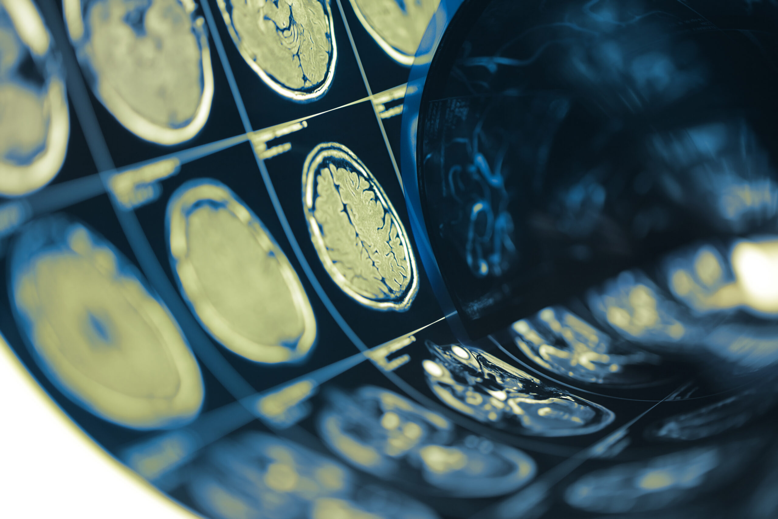
In the realm of optic neuritis diagnostics, radiological findings play a crucial role in delineating cases accurately. Utilizing advanced imaging techniques, particularly magnetic resonance imaging (MRI), researchers analyzed the radiological features associated with the false-positive cases defined by the 2022 optic neuritis criteria. The imaging results provided significant insights into the structural integrity of the optic nerves and any other relevant central nervous system (CNS) findings.
The imaging studies predominantly revealed variations in signal intensity and the presence or absence of distinct lesions, which are often pivotal in confirming optic neuritis. Many patients presented with normal findings on MRI, contradicting the expectations for a definitive diagnosis, while others exhibited non-specific white matter lesions frequently associated with migraines or other demyelinating conditions rather than optic neuritis itself.
Table 2 outlines the key radiological findings observed in the false-positive cohort, alongside their prevalence:
| Radiological Finding | Patients (n) | Percentage (%) |
|---|---|---|
| Normal MRI | 20 | 33.33 |
| Non-specific white matter lesions | 30 | 50 |
| Active lesions (demyelination) | 10 | 16.67 |
| Optic nerve enhancement | 5 | 8.33 |
| Other CNS findings | 8 | 13.33 |
A notable observation was the enhancement of the optic nerve in a minority of cases, which can often lead to misdiagnosis. However, in these false-positive cases, optic nerve enhancement did not correlate with clinical findings typical of optic neuritis but was rather isolated and not substantiated by the patients’ symptomatology.
The presence of non-specific white matter lesions was common, highlighting the challenges in distinguishing these from true inflammatory lesions associated with optic neuritis. These findings necessitate a nuanced approach to interpretation, taking into account the patient’s clinical presentation and history.
Moreover, the investigation unveiled that a significant percentage of patients had other CNS findings unrelated to optic neuritis. These included anomalies associated with benign conditions, which could further complicate diagnosis if not scrutinized thoroughly. Thus, comprehensive imaging alongside careful clinical evaluations is essential in preventing misdiagnoses that could lead to inappropriate treatments or interventions.
As this analysis demonstrates, a thorough understanding of radiological features associated with optic neuritis is pivotal, especially in cases presenting atypical symptoms or MRI findings. Enhanced diagnostic precision will ultimately lead to better-targeted therapies and improved patient outcomes.
Comparison with Established Criteria
Future Directions
The emerging insights from the analysis of false-positive cases classified by the 2022 optic neuritis criteria underline the necessity for a reevaluation of diagnostic frameworks and the incorporation of more refined methodologies for accurate classification. As the field of neuro-ophthalmology evolves, several critical avenues for future research and clinical practice can be identified to address the challenges currently faced.
Firstly, there is a pressing need for the development of an integrated diagnostic approach that combines clinical evaluation with advanced imaging techniques and biomarker identification. Current MRI standards may not sufficiently differentiate between true optic neuritis and other conditions presenting with similar symptoms. Therefore, future studies should focus on enhancing the sensitivity and specificity of MRI protocols, possibly through the inclusion of functional imaging and advanced diffusion-weighted imaging that can better characterize the nature of lesions found in the optic nerves.
In addition, the role of cerebrospinal fluid (CSF) analysis could be more prominently utilized to identify specific biomarkers that delineate optic neuritis from mimicking conditions. Studies have suggested that the presence of certain oligoclonal bands or inflammatory markers in the CSF can provide critical information that complements imaging findings and clinical symptoms.
Moreover, expanding the demographic and clinical diversity of study populations in future research can elucidate variations in optic neuritis presentations. This includes more extensive examination of pediatric cases and individuals with atypical risk factors, such as a history of migraine or other neurological disorders, which may predispose them to false-positive classifications.
Table 3 summarizes potential research directions for addressing the complexities surrounding optic neuritis diagnostics:
| Research Direction | Focus Area | Potential Outcomes |
|---|---|---|
| Integrated Diagnostics | Combining clinical, imaging, and biomarker assessments | Improved classification accuracy |
| Advanced MRI Techniques | Implementation of functional and diffusion-weighted imaging | Better differentiation of lesions |
| Cerebrospinal Fluid Analysis | Identification of inflammatory and oligoclonal biomarkers | Enhanced diagnostic precision and understanding of pathophysiology |
| Diverse Population Studies | Inclusion of broader demographic variables | Comprehensive insights into false-positive cases |
Furthermore, ongoing education and training for clinicians are essential to improve recognition of the varying manifestations of optic neuritis and its mimics. As knowledge of neuro-inflammatory conditions expands, the clinical community must mirror this growth by updating training modules that reflect current research findings and emphasize the importance of comprehensive history-taking and clinical examination.
Continued interdisciplinary collaboration among neurologists, radiologists, and ophthalmologists will further enhance patient care. Regular case discussions and interdisciplinary conferences can serve as platforms to share insights and refine diagnostic criteria based on collective experiences and evolving literature.
Ultimately, a refined, evidence-based approach to the diagnosis and management of optic neuritis and its false-positive cases will likely pave the way for enhanced patient outcomes, fostering a more accurate understanding of this complex condition.
Future Directions
Comparison with Established Criteria
The evaluation of false-positive cases classified by the 2022 optic neuritis criteria necessitates a thorough comparison with established diagnostic criteria, notably the previously utilized criteria such as the McDonald criteria and the criteria proposed by the Optic Neuritis Study Group. These frameworks have long dictated the diagnostic landscape for optic neuritis, yet the emergence of false-positive classifications highlights potential discrepancies and areas for refinement.
One primary difference observed in false-positive cases is the clinical presentation that often diverges from classic optic neuritis. Under the McDonald criteria, optic neuritis is traditionally characterized by acute onset of vision loss, pain with eye movement, and supportive MRI findings, including enhancement of the optic nerve. However, samples drawn from the recent analyses suggest that patients classified as false positives may present with ambiguous symptoms or atypical radiological findings, potentially leading to misclassification.
Table 4 below compares key features highlighted in false-positive cases against those outlined by established criteria:
| Feature | False-Positive Cases | Established Criteria (e.g., McDonald) |
|---|---|---|
| Onset of symptoms | Gradual or vague; may mimic other conditions | Acute onset of vision loss |
| Pain with eye movement | Present but less common | Commonly reported |
| Imaging findings | Normal MRI or nonspecific changes | Optic nerve enhancement; specific lesion identification |
| Demographic trends | Increased variability, including atypical cases | Predominantly young females |
Furthermore, the established criteria focus heavily on MRI findings, which in many cases do not decisively indicate active inflammation in false-positive cohorts. For instance, while the enhancement of the optic nerve is expected in confirmed optic neuritis, in about 8.33% of false-positive cases, such enhancement does not correlate with clinical symptoms indicating optic neuritis, thereby complicating diagnostic pathways.
Given these discrepancies, a revisitation of existing diagnostic criteria is warranted. Enhanced criteria could potentially include a broader scope of symptoms and consider alternative diagnostic pathways or tests that may help distinguish true optic neuritis from other neurological conditions presenting similarly. This may also involve reevaluating the role of ancillary diagnostic techniques, such as visual evoked potentials or the introduction of novel markers, that could serve to refine the classification framework.
Comparative studies involving longitudinal data on visual outcomes in patients classified under the new criteria as compared to established ones could provide invaluable insights and uncover patterns that merit incorporation in future criteria enhancements. The ultimate goal is to create a diagnostic protocol that minimizes misclassification rates while optimizing treatment regimens for patients.