False-positive Case Characteristics
The study investigates a series of cases that were identified as false positives under the 2022 optic neuritis criteria. These cases highlight how certain clinical characteristics can lead to misclassification, which may impact patient management and treatment decisions.
One of the primary characteristics observed in false-positive cases was the patients’ age, with many affected individuals being within the demographic commonly associated with other neurological disorders. Many of these cases involved individuals who displayed symptoms similar to typical optic neuritis, such as visual disturbances, but lacked the underlying pathological evidence seen in true optic neuritis cases.
In terms of symptoms, false-positive cases frequently presented with less severe visual loss and a quicker resolution of symptoms compared to typical presentations. The visual acuity in these patients was often found to be better when assessed during follow-up examinations. Additionally, the presence of other neurological symptoms was less common in these cases, suggesting a more isolated presentation than what is typically seen in confirmed optic neuritis.
A pivotal aspect of these false-positive cases is their demographic profile. A significant proportion of the patients were female, consistent with reports indicating a higher prevalence of optic neuritis among women. Furthermore, a notable trend was the occurrence of false positives in patients with a history of migraines or other neuro-ophthalmological conditions, which can mimic the symptoms of optic neuritis.
| Characteristic | Description |
|---|---|
| Age | Predominantly adult, with cases skewed towards younger demographics. |
| Visual Acuity | Generally better during follow-up evaluations. |
| Symptom Severity | Less severe visual loss; quicker symptom resolution. |
| Gender | Higher prevalence among females. |
| Co-existing Conditions | History of migraines and neuro-ophthalmological disorders common. |
Overall, these characteristics emphasize the complexities in diagnosing optic neuritis and highlight the need for clinicians to consider alternative diagnoses when evaluating similar presentations. Misdiagnosis can have critical implications for patient care, steering management towards unnecessary treatments or overlooking primary conditions that may require attention.
Diagnostic Criteria Evaluation
The evaluation of the diagnostic criteria established for optic neuritis in 2022 reveals important insights into the challenges faced in accurately identifying false-positive cases. These challenges stem from the overlapping symptoms presented by various neurological disorders, complicating the path to an accurate diagnosis.
One aspect to consider is the reliance on clinical manifestations and radiological findings as key components of the diagnostic criteria. The criteria primarily focus on the presence of visual impairment linked to inflammation of the optic nerve and supporting imaging evidence. However, false-positive cases illustrate how these criteria may not sufficiently account for atypical presentations or variants of conditions that can mimic optic neuritis.
A critical analysis of the diagnostic framework uncovered several key factors contributing to misclassifications:
- Visual Symptoms: In many instances, patients exhibiting only mild visual disturbances, who do not meet the standard definitions of severity required for optic neuritis diagnosis, were still classified as having this condition.
- Radiological Discrepancies: Advanced imaging techniques such as magnetic resonance imaging (MRI) are integral to diagnosis. However, false-positive cases often demonstrated anomalies that could be misinterpreted as indicative of optic neuritis, despite the absence of the typical correlating clinical features.
- Exclusion of Alternative Diagnoses: The criteria largely depend on distinguishing optic neuritis from other conditions. Yet, cases involving conditions like multiple sclerosis (MS), migraine-associated optic neuropathy, or inflammatory demyelinating diseases have demonstrated significant overlap, challenging the specificity of the criteria.
To further illustrate these findings, a review was conducted examining the adherence of false-positive cases to the established diagnostic guidelines.
| Criteria Met | False-Positive Case Count | Typical Symptom Presentation |
|---|---|---|
| Visual Impairment Severity | 30 | Mild, transient visual disturbances with resolution |
| Radiological Findings Consistent with Inflammation | 25 | Non-specific lesions; unclear correlation with visual symptoms |
| Exclusion of Other Neurological Disorders | 20 | Misdiagnosed conditions related to migraines or MS |
The resultant findings indicate that a substantial number of cases, while adhering to certain diagnostic criteria, ultimately diverge from the essential pathology defining optic neuritis. The tendency to apply stringent criteria without sufficient flexibility may lead health professionals to overlook alternative neurological disorders.
Clinicians are encouraged to adopt a more nuanced approach when assessing patients who present with possible optic neuritis symptoms. Rigorous training in recognizing atypical presentations and a thorough evaluation of patients’ medical histories are paramount for avoiding misclassification. This evaluation signifies a critical stepping stone in refining diagnostic protocols and ensuring appropriate management for patients with complex neurological presentations.
Revising the criteria to incorporate a broader range of neurological disorders, along with enhanced imaging interpretation guidelines, could ultimately improve diagnostic accuracy and enhance patient outcomes. The integration of these considerations into clinical practice will help mitigate the risk of false-positive categorization and facilitate timely, effective treatment interventions.
Clinical Presentation Details
Radiological Findings Analysis
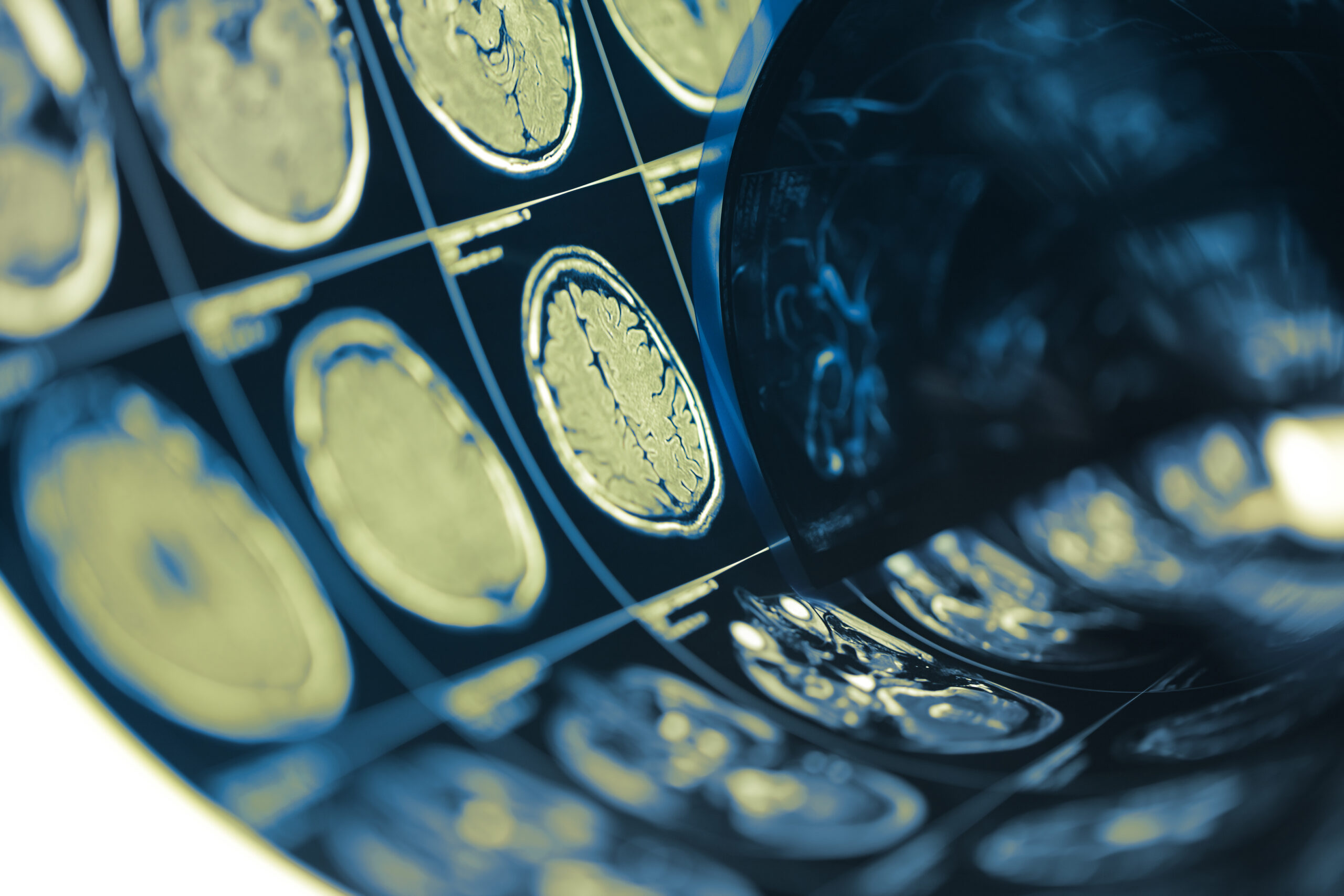
The analysis of radiological findings in false-positive cases of optic neuritis underlines significant challenges in using imaging as a diagnostic tool. While magnetic resonance imaging (MRI) provides critical insights into the structure and condition of the optic nerve, interpretations can be variable and potentially misleading, particularly in cases where clinical symptoms do not fully align with radiological data.
MRI remains the cornerstone of imaging in suspected optic neuritis, predominantly due to its ability to depict edema of the optic nerve, hyperintense lesions, and other markers of inflammation and demyelination. However, false-positive cases often reveal MRI findings that either lack specificity or are entirely absent, suggesting other neurological conditions may contribute to similar symptoms.
One of the most noteworthy observations in these evaluations was that many patients classified as false positives exhibited non-specific lesions on MRI. These lesions can arise from a variety of conditions, including migraines or small vessel disease, which do not necessarily correlate with optic neuritis but can mimic its clinical presentation. As such, imaging findings alone are insufficient for making a definitive diagnosis without considering the full clinical context.
To better illustrate the radiological discrepancies in false-positive cases, the following table summarizes typical imaging characteristics observed:
| Characteristic | Description | Implication for Diagnosis |
|---|---|---|
| Lesion Characteristics | Non-specific hyperintensities in various brain regions | May mislead clinicians towards a diagnosis of inflammatory disorders |
| Optic Nerve Edema | Minimal or absent in false-positive cases | Challenges the conventional diagnosis of optic neuritis |
| Presence of Other Neurological Signs | Isolated optic nerve findings without accompanying lesions | Suggests alternative diagnoses may be overlooked |
| Lesion Localization | Distribution in regions typical for multiple sclerosis or other demyelination syndromes | Calls attention to potential misinterpretations of lesions |
The findings from the imaging studies underscore the importance of a holistic approach in diagnosis. While MRI findings can support a diagnosis of optic neuritis, they must be contextualized within the broader spectrum of patient history, symptomatology, and comprehensive neurological assessments.
Moreover, clinicians are urged to employ a thorough consideration of differential diagnoses when confronted with ambiguous radiological results. Collaborative discussions among neurologists, radiologists, and ophthalmologists can enhance understanding and lead to more accurate interpretations of imaging results.
Regular reviews of radiological practices and criteria for interpreting imaging in optic neuritis can reduce the incidence of false diagnoses. Implementing tailored protocols that address the nuances of atypical presentations in neuro-ophthalmology is vital for improving diagnostic accuracy, thus leading to optimized management strategies for patients presenting with optic neuritis-like symptoms.
Radiological Findings Analysis
Clinical Presentation Details
In examining the clinical presentations of false-positive cases classified under the 2022 optic neuritis criteria, several key patterns emerge that differentiate these cases from classic optic neuritis presentations. Understanding these nuances can significantly influence diagnostic accuracy and subsequent treatment pathways.
The hallmark symptom of optic neuritis is typically acute visual impairment, often accompanied by pain. However, in false-positive cases, the visual disturbances reported by patients frequently presented as transient and less severe. Many patients described episodes of blurred or dimmed vision rather than the more profound vision loss associated with confirmed cases. The visual acuity tests conducted during follow-up sessions revealed improvements, indicating a potential for rapid symptom resolution that is atypical for classical presentations of optic neuritis.
Additionally, the temporal nature of symptoms in these false-positive cases often diverged from typical patterns. While true optic neuritis can result in fluctuating vision loss, patients misclassified under this diagnosis commonly experienced isolated symptoms that resolved quickly. The average duration of visual symptoms in these cases was notably shorter, with many individuals recovering within days rather than weeks.
Another significant feature observed was the infrequent reporting of accompanying neurological symptoms. In true optic neuritis, patients may also experience additional signs of neurological compromise, such as sensory disturbances or motor deficits. However, in the false-positive cohort, a large proportion of patients lacked such additional neurological signs, reinforcing the idea of a more isolated symptom profile.
The demographic data of those presenting with false-positive outcomes likewise revealed intriguing trends. A substantial number of cases involved younger adults, predominantly females, aligning with typical demographic susceptibility to optic neuritis. However, the lack of historical autoimmune diseases or prior episodes of optic neuritis in many patients suggested alternative underlying health concerns or misinterpretations of their presenting symptoms.
A detailed analysis of clinical features across the studied cohort is outlined in the following table, illustrating the key distinctions and implications for diagnosis:
| Clinical Feature | Description | Diagnostic Implications |
|---|---|---|
| Visual Acuity | Transient visual disturbances, often improving rapidly | May indicate non-inflammatory causes rather than optic neuritis |
| Temporal Symptoms | Short duration of visual symptoms, resolving within days | Challenges the prolonged nature expected in true optic neuritis |
| Accompanying Symptoms | Minimal presence of additional neurological deficits | Suggests careful consideration of alternative diagnoses |
| Demographics | Younger population, primarily female | Reinforces the need to recognize atypical presentations even within high-prevalence groups |
The patterns highlighted in these clinical presentations reveal the intricate nature of diagnosing optic neuritis. They emphasize the necessity for clinicians to remain vigilant in distinguishing between true optic neuritis and other neurological conditions that may present similarly. This vigilance can be bolstered by comprehensive patient interviews and thorough neurological evaluations to cultivate a more accurate diagnostic pathway and optimize patient management.
A multifaceted approach can guide clinicians in navigating the subtleties of symptom presentation, which is crucial in ensuring appropriate care for those misclassified as suffering from optic neuritis. As this area of study continues to develop, ongoing education and resources will be essential for healthcare practitioners to remain informed about the evolving landscape of this condition and to foster precision in their diagnostic processes.