Study Overview
The research aimed to investigate the structural changes that occur in the brain due to varying impact forces, using post-mortem human specimens. Recognizing the potential implications for understanding traumatic brain injury (TBI), this study specifically focused on how these deformations could enhance our knowledge of brain pathophysiology. Researchers sought to methodically analyze how different levels of mechanical pressure affect the brain’s anatomy, aiming to establish a correlation between injury severity and the resulting morphological changes within the cerebral structure.
This exploration was prompted by the need for more precise metrics in assessing brain trauma, especially in forensic contexts and clinical settings. By utilizing advanced imaging techniques and biomechanical analysis, the study endeavored to create a comprehensive overview of intracranial deformation, thereby contributing to a deeper understanding of TBI and the complex responses of brain tissue to external forces.
The selection of specimens was crucial, ensuring that a diverse representation of post-mortem cases was included to reflect variability in anatomical and physiological factors. Additionally, the research incorporated a range of impact conditions to better emulate real-world scenarios that could lead to traumatic brain injuries, allowing for a thorough examination of how impact variables correlate with specific brain deformation outcomes.
Methodology
The study meticulously utilized a combination of advanced imaging techniques and biomechanical assessments to investigate the brain deformation resulting from controlled impact scenarios. Post-mortem human specimens were selected from a diverse pool, encompassing a range of ages, genders, and pre-existing health conditions, which provided a more representative sample of the human population. This diversity ensured that various anatomical and physiological factors were accounted for during analysis, which is critical in studying the effects of trauma on the brain.
Each specimen underwent a series of carefully controlled impact tests. These tests were designed to replicate a spectrum of force levels that might occur during real-life traumatic events, such as falls or vehicle collisions. Researchers employed a drop tower apparatus, which allowed for precise control over the drop height and, consequently, the impact force applied to the specimens. The forces were calibrated to generate impacts classified as low, moderate, and severe, mimicking different clinical scenarios associated with traumatic brain injury.
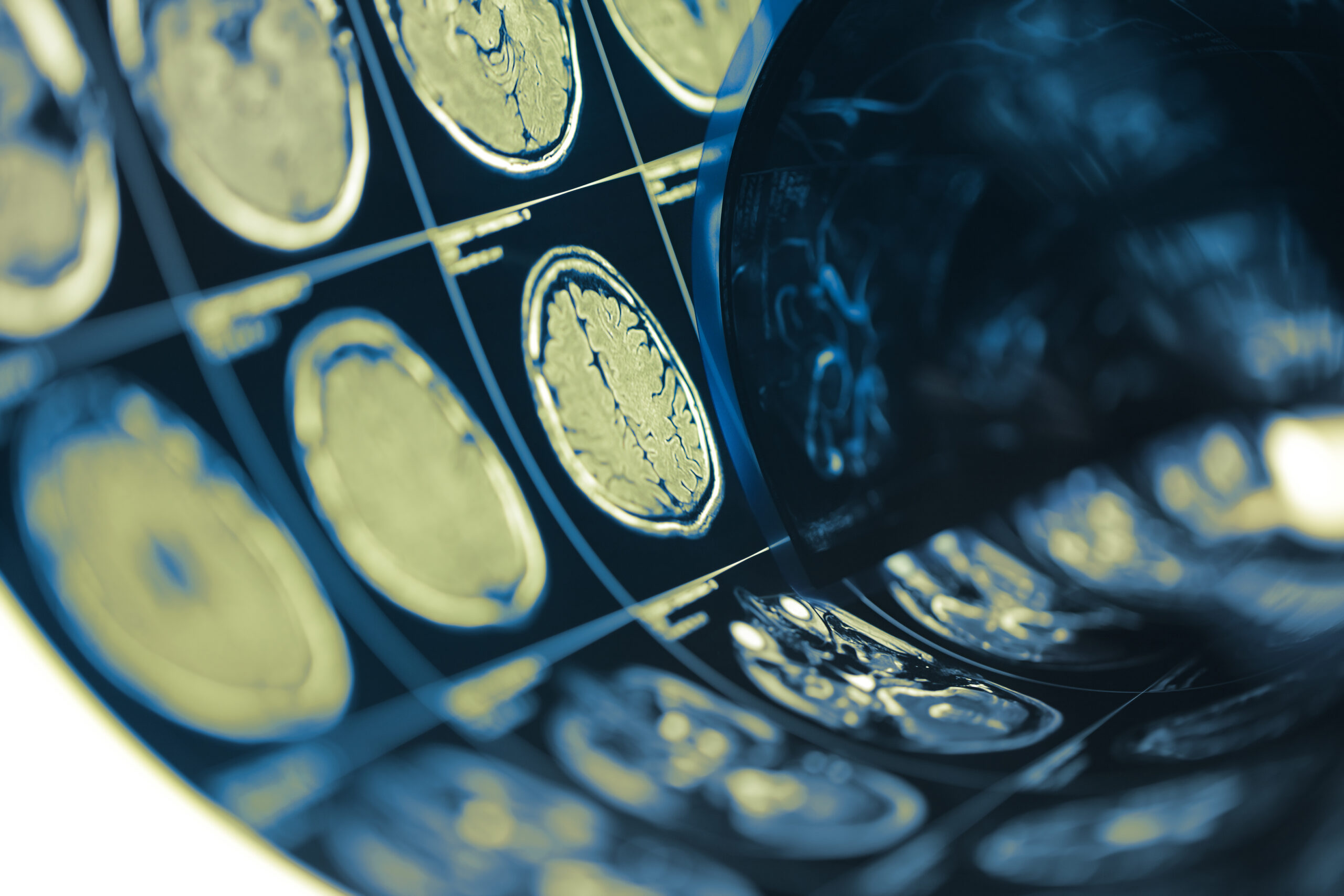
Following the application of impact forces, high-resolution imaging techniques, including Magnetic Resonance Imaging (MRI) and Computed Tomography (CT), were employed to capture the anatomical changes in the brain. These imaging modalities provided detailed three-dimensional visualizations of the cerebral structures, enabling researchers to observe subtle alterations in morphology and volume. The images were subjected to sophisticated analytical software, aimed at quantifying deformations and identifying specific areas of interest, such as cortical and subcortical regions.
Additionally, the study incorporated finite element modeling (FEM) as part of the biomechanical analysis. This computational method allowed researchers to simulate the mechanical behavior of brain tissue under stress, providing insight into the mechanics of deformation and strain distribution throughout the brain. By correlating the deformation patterns observed in imaging studies with FEM outputs, the researchers were able to draw robust connections between the severities of impact and the resultant structural changes.
Data collection also included the documentation of relevant clinical histories of the specimens, enabling researchers to consider factors such as previous head trauma, neurological conditions, and other health variables that might influence the susceptibility of brain tissues to deformation. These comprehensive datasets facilitated a more robust analysis of how individual differences might affect brain response to impact.
Statistical methods were employed to analyze the relationships between impact forces, demographic factors, and observed deformations. Various models, including regression analyses, were utilized to explore the significance of the findings and to identify potential predictive markers that could indicate the risks of TBI related to specific impact conditions. This integrative approach, combining experimental, clinical, and computational strategies, enhanced the study’s validity and reliability, ultimately providing a clearer understanding of the complex interplay between mechanical forces and brain injury.
Key Findings
The study yielded several pivotal insights into the relationship between impact forces and brain deformation. First and foremost, it was established that increased impact intensity directly correlates with the magnitude of structural changes observed in brain specimens. Specifically, the data revealed that specimens subjected to severe impacts exhibited pronounced deformations compared to those exposed to moderate or low forces. This correlation supports existing hypotheses linking higher impact forces to elevated risks of severe traumatic brain injury (TBI).
High-resolution imaging analyses demonstrated that key areas of the brain, particularly the cortical and subcortical regions, displayed varying degrees of deformation contingent upon the level of impact. The quantitative measurements indicated that areas associated with cognitive and motor functions were particularly vulnerable to deformation when subjected to high force impacts, suggesting a potential pathway for understanding the mechanisms underlying cognitive decline and neurodegeneration post-injury.
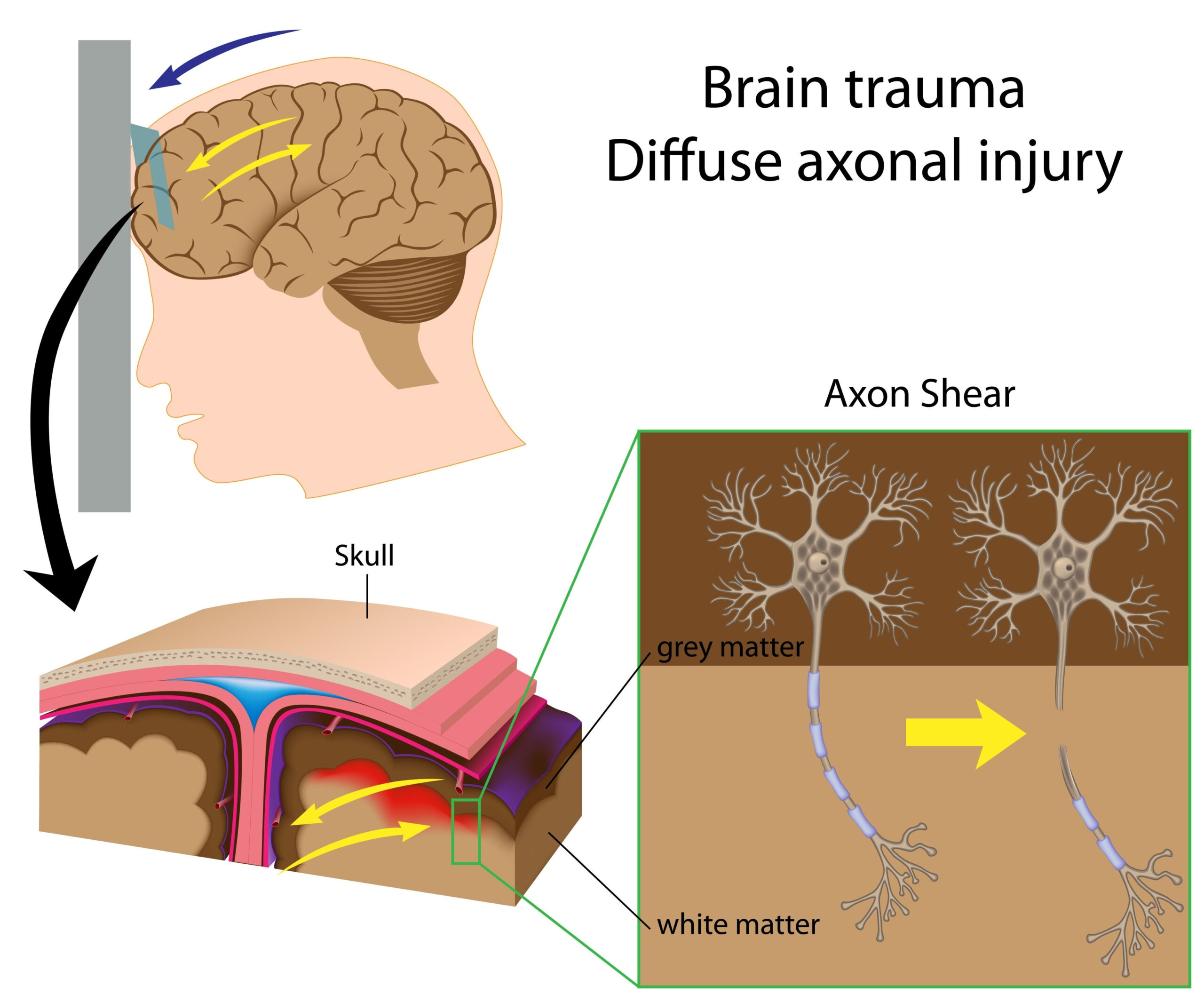
Moreover, the integration of finite element modeling provided critical insights into the biomechanical responses of brain tissue during impact. The computational simulations indicated that different regions of the brain experienced distinct stress and strain patterns based on the localized forces applied. For instance, the simulations predicted that while the surface layers of the cortex may yield to shear forces, deeper structures, such as the white matter tracts, exhibited tensile stresses that could lead to subtle but significant disruptions over time.
Another significant finding was the impact of demographic factors, such as age and gender, on brain response to trauma. Older specimens showed a tendency for increased susceptibility to deformation at lower impact forces compared to younger counterparts. This suggests that age-related changes in brain structure, such as reduced tissue elasticity and increased stiffness, may play a critical role in the degree of deformation experienced during traumatic events. Furthermore, gender differences were noted, with male specimens demonstrating greater resilience to deformation, potentially reflecting inherent biological variations in brain composition and structure.
The study also highlighted the influence of pre-existing health conditions on brain deformation outcomes. Specimens with a history of previous head trauma displayed exacerbated deformation patterns even at lower impact forces, underscoring the cumulative effects of multiple injuries and the importance of considering individual medical histories when assessing susceptibility to TBI.
Importantly, statistical analyses confirmed the robustness of these findings, revealing notable patterns that emerged consistently across different demographic and clinical groups. By outlining specific predictive markers linked to the severity of brain deformation based on impact conditions, this research paves the way for enhanced assessing strategies in both clinical and forensic settings. Such markers could potentially guide prognosis and treatment plans for individuals at risk of sustaining neurotrauma.
These findings significantly advance our understanding of how varying impact forces influence brain structure, shedding light on critical factors that might inform prevention and intervention strategies for traumatic brain injuries. The intricate interplay among impact severity, demographic profiles, and individual health conditions revealed in this study emphasizes the need for continued research to develop comprehensive approaches to both the assessment and management of TBI.
Strengths and Limitations
The strengths of this study lie in its methodological rigor, comprehensive data collection, and multifaceted approach to analyzing brain deformation due to mechanical impacts. A key advantage is the utilization of post-mortem human specimens, allowing for direct observation of the brain’s physiological responses under controlled conditions that closely mimic real-world traumatic events. This provides a significant improvement over previous studies that might have relied heavily on animal models or theoretical simulations that do not account for human anatomical variability.
Another strength is the incorporation of advanced imaging techniques such as MRI and CT, which offer high-resolution visualizations of brain structures and allow for nuanced assessments of morphological changes. These imaging modalities, combined with the use of finite element modeling, enable a deeper understanding of the biomechanics of brain tissue under stress, facilitating the identification of distinct deformation patterns across different regions of the brain. This integrative approach strengthens the validity of the findings and enhances the ability to draw meaningful conclusions about the impact of forces on various brain structures.
Furthermore, the focus on a diverse population of specimens, with careful documentation of demographic factors and previous health histories, amplifies the study’s significance. The inclusion of age and gender as variables provides valuable insights into how biological differences may influence brain vulnerability to trauma. By exploring the interplay between individual characteristics and susceptibility to brain deformation, the research contributes to a more personalized understanding of traumatic brain injuries and underscores the importance of tailoring prevention strategies to specific populations.
However, this study does present certain limitations that warrant consideration. One notable constraint is the reliance on post-mortem specimens, which, while offering unique insights, may be subject to various confounding factors, including the effects of post-mortem changes on brain tissues and potential biases in specimen selection. Additionally, while efforts were made to ensure a wide-ranging sample, the consistency of findings across all demographic categories remains to be confirmed in future research using larger and more varied cohorts.
Another limitation is the controlled nature of the impact tests, which, although meticulously executed, may not fully replicate the complexities of real-life scenarios where multiple factors—such as angling of impact, rotational forces, and varying tissue health—could contribute significantly to injury outcomes. These factors, if not accounted for in the current dataset, could limit the generalizability of the results to everyday situations involving traumatic brain injuries.
Moreover, the integration of finite element modeling, while powerful, relies on specific assumptions and parameters that must be carefully validated against empirical observations. Any inaccuracies in the modeling could potentially affect the interpretation of the biomechanical responses observed in the specimens, necessitating further validation through longitudinal studies or the application of alternative modeling techniques.
While this study presents a robust framework for understanding the relationship between impact forces and brain deformation, awareness of its limitations is crucial for accurately interpreting the findings. Continued research with an emphasis on expanding sample diversity and exploring the complexities of real-world impact scenarios will be essential for advancing knowledge in this critical area of traumatic brain injury research.