Study Overview
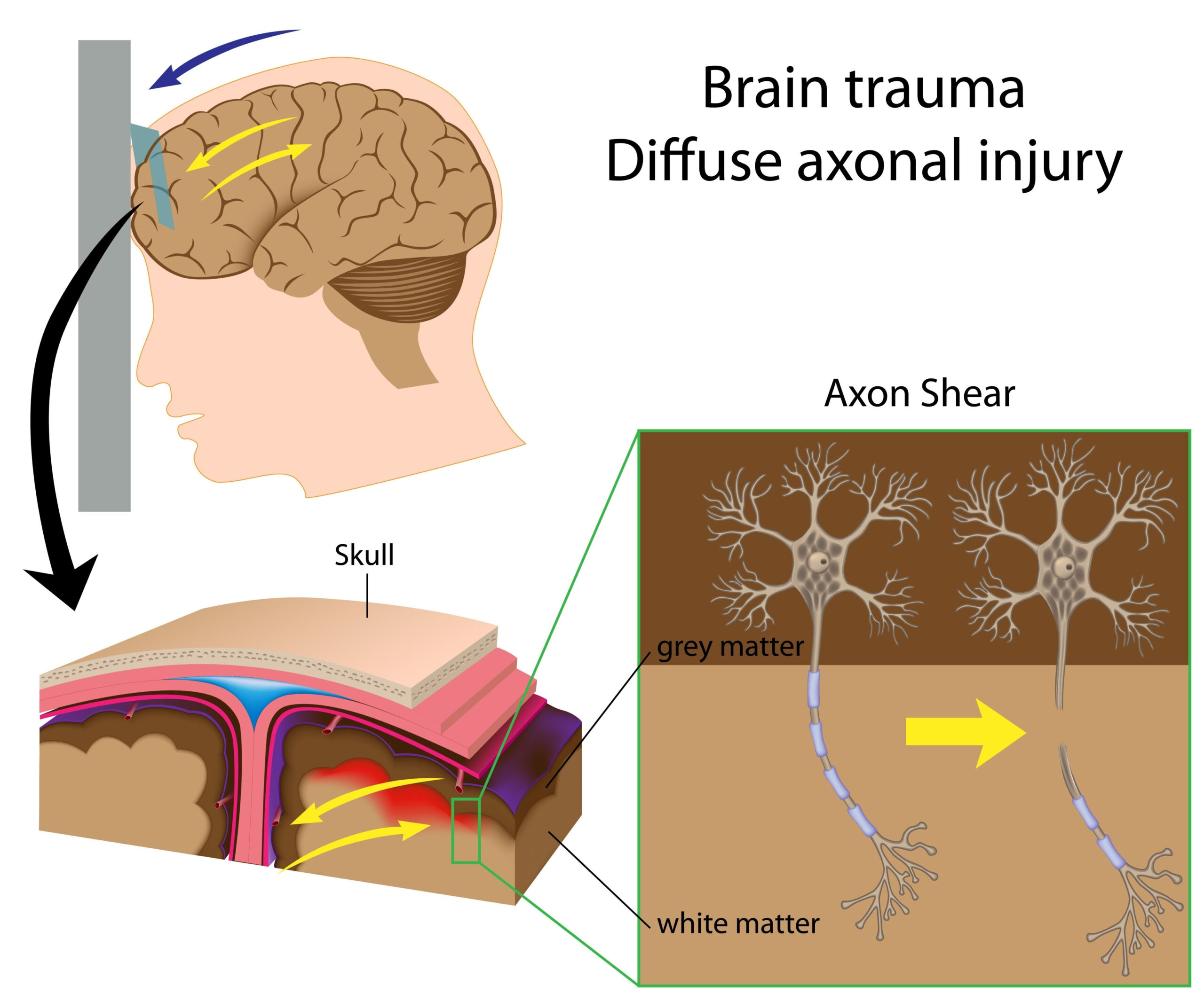
The research investigates the complex interactions between inflammation and endocrine responses following combined burn and blast injuries in a model that simulates a trauma environment. This study primarily aims to elucidate how these injuries impact early brain responses, which are crucial for understanding subsequent neurological and systemic outcomes. The injury model utilized underscores the dual challenges posed by thermal and mechanical traumas, which can significantly alter physiological responses compared to isolated injuries.
The study employs a comprehensive approach, combining both in vivo experimentation and biochemical assessments to capture the multifaceted nature of the responses elicited by such complex trauma. It focuses on examining various biomarkers of inflammation and hormone signaling pathways that are activated during the early post-injury phase. By using advanced technologies and analytical methods, the researchers aim to provide insights into the crosstalk between different organ systems—specifically how signals from immune and endocrine systems influence brain function in the setting of acute trauma.
The significance of this research arises from the necessity to develop targeted therapeutic strategies that not only address the immediate physical damage caused by burn and blast injuries but also consider the broader implications of these injuries on brain health and function. These insights may consequently lead to improved management protocols for patients experiencing multiple organ injuries, ultimately enhancing recovery outcomes. This study stands out for its potential to bridge the gap between basic scientific understanding and clinical application, offering a promising direction for future investigations in trauma medicine.
Methodology
The investigation employs a robust methodological framework designed to capture the intricate physiological and biochemical responses following combined burn and blast injuries. Utilizing an established animal model, the researchers implemented controlled trauma to mimic the physical and emotional stresses experienced in a real-world scenario. This approach allows for a close examination of how these dual injuries impact the body’s regulatory systems, particularly regarding inflammation and endocrine signaling.
The study commenced with the selection of a suitable animal model, specifically adult male rodents, which were subjected to standardized burn and blast protocols. The burn component involved applying heat to induce thermal injury, while the blast injury was simulated using a shock wave generator. These procedures were carefully calibrated to ensure uniformity and reproducibility, critical for deriving meaningful data. Following the injuries, the subjects were closely monitored for various physiological parameters.
In parallel, the researchers collected serum samples at multiple time points post-injury to analyze a range of biomarkers associated with inflammation and hormone levels. Key cytokines and hormones were measured, including interleukins, tumor necrosis factor-alpha (TNF-α), cortisol, and catecholamines. These biomarkers provide critical insights into the activation of the immune response and the endocrine systems, which are pivotal in modulating brain function and overall recovery.
Histological analyses were conducted on brain tissues to assess the extent of cellular and structural changes resulting from the injuries. Techniques such as immunohistochemistry allowed for the visualization of specific inflammatory markers within brain tissue, facilitating an understanding of the spatial distribution of inflammatory responses. Additionally, molecular techniques were applied to evaluate gene expression profiles related to inflammation, neuroprotection, and hormonal responses, enriching the understanding of the underlying mechanistic pathways triggered by combined injuries.
Data analysis involved a combination of statistical methods to compare pre-injury and post-injury states across different groups, ensuring that any observed differences could be attributed to the traumatic events rather than natural variability. The intention behind this comprehensive methodology is to establish a clear connection between the biochemical responses and resultant neurological outcomes, thus contributing to a nuanced understanding of how multi-organ cross-talk occurs following traumatic injury.
Through this meticulous approach, the study aims to uncover pivotal insights into the interactions between the immune and endocrine systems in the early phases of combined burn and blast injuries. These findings not only enhance our biological understanding but also have the potential to influence clinical interventions aimed at mitigating adverse outcomes in trauma patients.
Key Findings
The investigation yielded several critical insights into the physiological alterations that occur following combined burn and blast injuries. The study found significant elevations in specific biomarkers, thereby highlighting the interplay between inflammatory responses and hormonal regulation in the brain during the acute post-injury phase.
One of the most pronounced findings was the marked increase in pro-inflammatory cytokines, including interleukin-6 (IL-6) and tumor necrosis factor-alpha (TNF-α), in serum samples collected shortly after the injuries. These cytokines are known mediators in the inflammatory response and play an essential role in the communication between the immune system and the central nervous system. Notably, the trajectory of these cytokines was closely linked to the severity of the injuries, suggesting that the systemic inflammatory response directly influences the extent of neural disruption.
Furthermore, analyses revealed that hormone levels, particularly cortisol and catecholamines such as epinephrine and norepinephrine, were significantly elevated post-injury. The rise in cortisol, a stress hormone, correlates with the body’s attempt to manage the shock resulting from trauma. This hormonal response not only helps regulate metabolism and immune function but is also indicative of the body’s stress response mechanism, which may compound the inflammatory effects observed.
Histological evaluations of brain tissue indicated that the combined injuries led to pronounced neuroinflammatory changes, characterized by increased activation of astrocytes and microglia. These glial cells are integral to maintaining homeostasis within the central nervous system, yet their overactivation can lead to detrimental effects, including neuronal injury and apoptosis. Immunohistochemical staining provided evidence of increased expression of inflammatory markers specifically localized to areas of the brain associated with pain perception and cognitive functions, suggesting that such injuries may lead to long-term neurological deficits.
Molecular assessments uncovered alterations in gene expression related to neuroprotection and apoptosis. Notably, there was a notable increase in the expression of genes associated with inflammatory pathways, indicating that the brain’s defensive mechanisms are critically engaged in response to traumatic stressors. Conversely, genes associated with cellular protection showed variance in expression, signaling an adaptive yet potentially overwhelmed neuroprotective response in the setting of combined traumatic injuries.
Statistical analyses reinforced these findings, demonstrating significant differences in biomarker levels and gene expression profiles when contrasting pre-injury states with post-injury measurements. These results underscore the complexity of multi-organ interactions, emphasizing that the acute response to dual insults extends well beyond the localized injuries and has a profound influence on overall neurological health.
In summary, this study illuminates the intricate biochemical pathways activated by combined burn and blast injuries, revealing a robust inflammatory and endocrine response that profoundly impacts brain function. The documented changes not only emphasize the need for targeted therapeutic strategies in managing trauma but also highlight potential avenues for further research into the long-term effects of such traumatic injuries on neurological health and recovery.
Clinical Implications
The findings from this study underscore the necessity for an integrative approach to the management of individuals suffering from combined burn and blast injuries. Recognizing the interconnectedness of inflammatory and endocrine responses is essential for developing effective treatment protocols aimed at improving patient outcomes. As multi-organ injuries not only compromise physical health but also have critical ramifications for neurological functions, clinicians must adopt a multidisciplinary perspective when treating such injuries.
The pronounced elevation of pro-inflammatory cytokines, particularly IL-6 and TNF-α, indicates a robust inflammatory response that accompanies these traumatic events. Clinicians might consider the early assessment of these biomarkers as a valuable component of triage and prognostication. Early identification of heightened inflammatory states could prompt timely interventions, such as the administration of anti-inflammatory agents. Targeted therapies aiming to mitigate excessive inflammation could not only alleviate immediate symptoms but also potentially reduce the risk of long-term cognitive and emotional impairments.
Moreover, the observed hormonal fluctuations, including increased cortisol and catecholamines, elucidate a physiological stress response that necessitates careful monitoring. While these hormones are crucial for managing acute stress, prolonged elevation could exacerbate deleterious effects on neurological health. Clinicians should consider strategies to balance these endogenous stress responses, potentially employing pharmacological or psychological interventions that promote recovery while preventing the adverse outcomes associated with chronic stress.
Histological changes noted in the brain, characterized by activated astrocytes and microglia, serve as warning signs that further underscore the need for interventions focusing on neuroprotection. The potential for long-lasting neuroinflammatory changes suggests that monitoring cognitive functions and mental health post-injury should become routine practice. Interventions, which may include neuroprotective agents or rehabilitative therapies that target cognitive and emotional rehabilitation, could be crucial in mitigating the long-term consequences of such injuries.
Additionally, the alteration in gene expression profiles indicates that the brain’s ability to protect itself is not only impacted by the immediate response to trauma but may also reflect adaptations that require medical attention. This insight aligns with the growing recognition of personalized medicine, where treatments can be tailored based on an individual’s specific inflammatory and hormonal responses.
Ultimately, these findings advocate for enhanced clinical protocols that incorporate the assessment of both systemic inflammatory markers and hormonal profiles. Such integrative approaches promise to foster better recovery trajectories by addressing the biological underpinnings of trauma, leading to optimized rehabilitation and supportive care in clinical settings. By expanding the focus from immediate injury management to also encompass the broader systemic and neurological implications of trauma, healthcare providers can significantly improve the quality of care for individuals with complex injury profiles.