Impact of Blast-Induced Injuries on Brain Structure
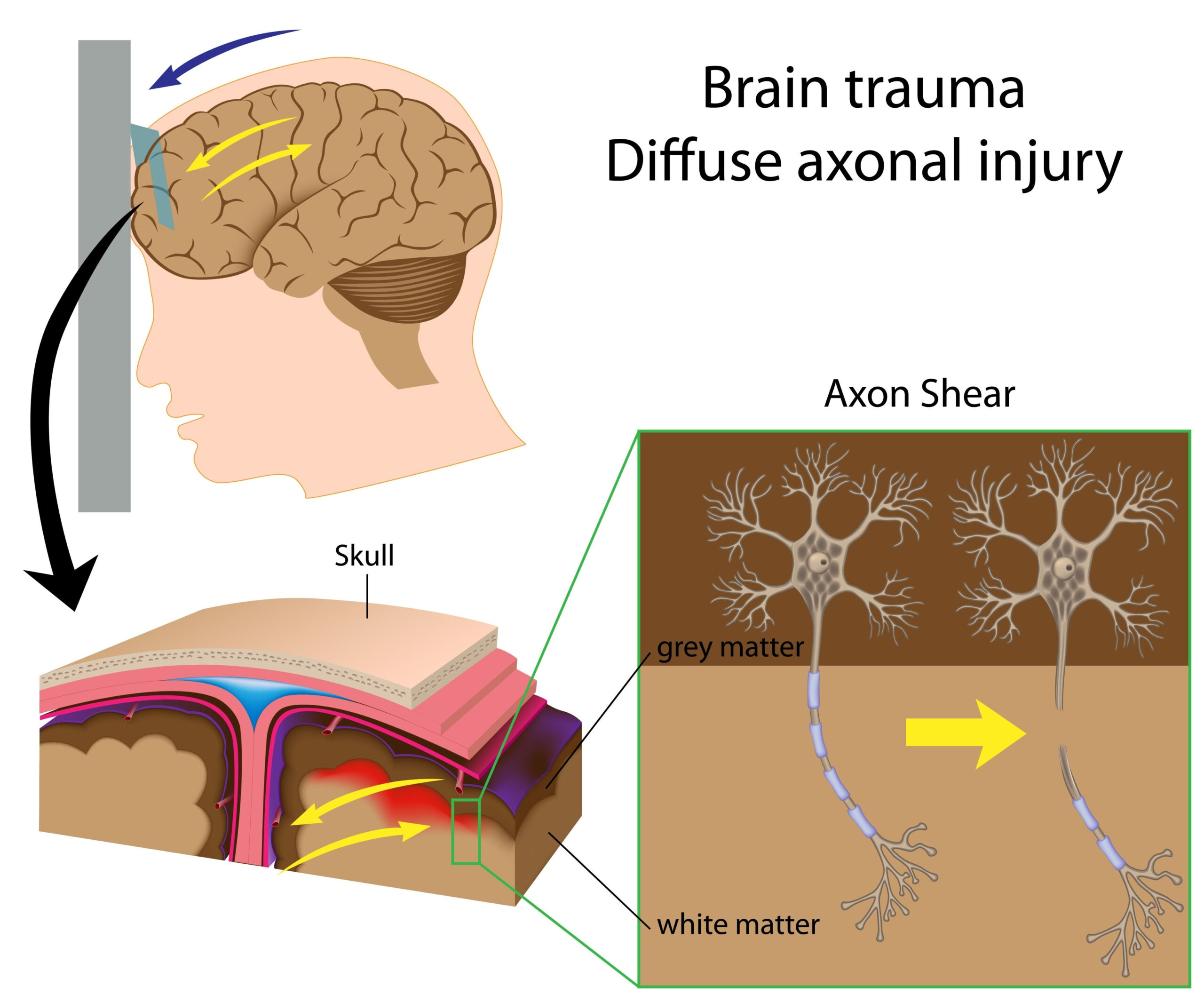
Blast-induced traumatic brain injury (TBI) significantly alters the structural integrity of the brain, particularly impacting vulnerable regions such as the hypothalamus. The hypothalamus plays a critical role in regulating numerous autonomic functions, and its damage can have cascading effects throughout the body. Research indicates that blast waves can lead to both primary and secondary injuries. Primary injuries are caused by the direct mechanical force of the blast, resulting in the displacement of brain tissue, which can cause physical damage to neurons and glial cells. This mechanical injury can lead to immediate disruptions in cellular function, contributing to neurodegeneration and inflammation.
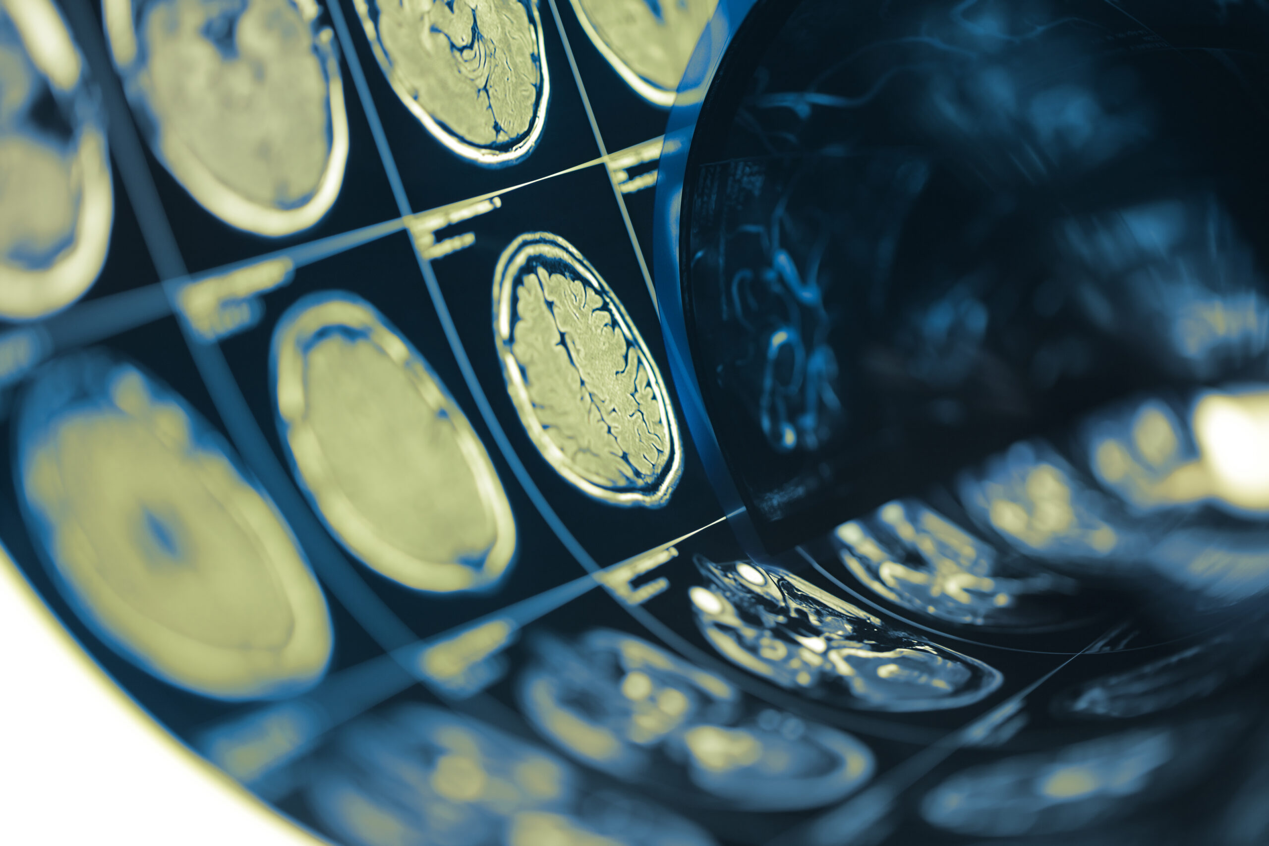
Secondary injuries may arise from the inflammatory response triggered by the initial trauma. Following the blast, the brain releases a variety of inflammatory cytokines and chemokines, which can exacerbate cellular damage and promote further neuroinflammation. Studies utilizing advanced imaging techniques like MRI have shown alterations in brain morphology, demonstrating reduced volume in impacted areas, including the hypothalamus, following exposure to blast forces. These changes in brain structure have been correlated with functional impairments, highlighting the significance of understanding the specific areas affected by blast-induced injuries.
Furthermore, animal models and post-mortem analyses provide compelling evidence that repeated exposure to blast can lead to progressive structural changes over time. Findings suggest that neuronal death and axonal injury become more pronounced with consecutive blasts, often resulting in chronic issues such as chronic traumatic encephalopathy (CTE). The hypothalamus, which governs critical hormonal functions and the body’s circadian rhythms, could exhibit altered neuroarchitecture that impacts its ability to maintain homeostasis under stress conditions elicited by blast exposures. Thus, examining the structural impact of blast-related injuries on the brain, especially the hypothalamus, is crucial for understanding their long-term effects on cognitive and physiological functions.
Experimental Design and Procedures
This study aimed to investigate the biochemical and structural effects of blast-induced traumatic brain injury (TBI) on the hypothalamus. A comprehensive approach was employed, utilizing both in vivo and in vitro methodologies to assess the direct impacts of blast exposure on brain tissue. Animal models were chosen for this research due to their relevance in mimicking human physiological responses to external forces.
The experiment involved a cohort of male and female rodents, which were selected to provide insights into potential sex differences in response to blast exposure. The animals underwent controlled blast simulations, where they were subjected to pressure waves generated by explosive materials. The specific parameters of the blast, including distance from the source and intensity, were standardized to ensure reproducibility. Careful monitoring was in place to assess immediate behavioral responses, which included changes in motor activities and exploratory behaviors.
Post-exposure, subjects were euthanized at various time points to collect brain tissues for histological and biochemical analyses. Histopathological examinations utilized techniques such as hematoxylin and eosin (H&E) staining, which allowed for visualization of structural changes in neuronal cells and glial populations. The hypothalamus was specifically targeted for analysis due to its critical homeostatic functions, including regulation of endocrine signaling and autonomic responses.
In addition to structural assessments, biochemical markers of injury were evaluated. This included measuring levels of inflammatory cytokines and oxidative stress markers in hypothalamic tissue samples using enzyme-linked immunosorbent assays (ELISA). The intent was to pinpoint the neuroinflammatory processes that ensue following blast exposure and establish a correlation with observed structural changes. Neurotransmitter levels and metabolic changes were assessed through high-performance liquid chromatography (HPLC), providing a detailed profile of biochemical alterations in the hypothalamus.
To dissect the temporal dynamics of injury progression, tissues were collected at several intervals—immediately post-exposure, at 24 hours, one week, and four weeks after the blast. This longitudinal approach enabled researchers to identify both acute and chronic effects, mapping out the trajectory of neuronal damage and recovery within the hypothalamic region.
Statistical analyses were performed to compare results across different time points and treatment groups, employing multifactorial ANOVA to determine significant differences in structural and biochemical outcomes. The multifactorial design considered variations based on sex and specific blast exposure parameters, allowing for nuanced interpretations of the data.
This rigorous experimental design facilitated a thorough exploration of how blast-induced injuries affect the structure and biochemistry of the hypothalamus, ultimately guiding further studies into therapeutic interventions and long-term implications for individuals exposed to blast trauma.
Biochemical Changes in the Hypothalamus
Following blast-induced traumatic brain injury (TBI), the hypothalamus undergoes a series of significant biochemical alterations that can further compound the effects of mechanical damage. These changes manifest in various molecular pathways involving inflammation, oxidative stress, and neurotransmitter disruptions, all of which are crucial for maintaining the hypothalamus’s regulatory functions. The biochemical response to blast exposure initiates immediately and can persist well beyond the initial injury, contributing to long-term dysfunction.
One of the primary responses observed in the hypothalamus post-blast is an elevation in pro-inflammatory cytokines. Cytokines such as interleukin-6 (IL-6) and tumor necrosis factor-alpha (TNF-α) have been found to increase significantly within hours of exposure. These inflammatory markers can disrupt the normal balance of neuronal signaling, leading to altered homeostatic responses including disrupted hormonal regulation. For instance, increased IL-6 levels have been linked to disturbances in the hypothalamic-pituitary-adrenal (HPA) axis, affecting stress responses and potentially leading to disorders such as anxiety or depression in affected individuals.
Additionally, oxidative stress plays a pivotal role in the biochemical changes following blast injuries. The brain, particularly the hypothalamus, is highly susceptible to oxidative damage due to its rich lipid content and high metabolic rate. Post-blast, there is an increase in reactive oxygen species (ROS), which, coupled with the diminished antioxidant defenses, leads to lipid peroxidation and neuronal cell death. Elevated levels of malondialdehyde (MDA), a byproduct of lipid peroxidation, serve as a quantitative marker for oxidative damage, presenting a clear link between blast exposure and cellular injury.
Neurotransmitter systems also display significant alterations; for example, changes in levels of norepinephrine and serotonin have been documented following blast exposure. These neurotransmitters are integral to mood and cognitive functions, and disruptions can lead to a spectrum of neuropsychiatric conditions. High-performance liquid chromatography (HPLC) analyses have revealed these neurotransmitter imbalances, indicating a shift that may contribute to the behavioral and cognitive impairments often observed in individuals suffering from blast-related injuries.
Moreover, the hypothalamus’s metabolic activities, including glucose regulation and energy homeostasis, are notably affected post-injury. Insulin signaling pathways are compromised, which can induce a state of insulin resistance. Such metabolic dysregulation not only impacts energy balance but may also influence overall health outcomes, leading to obesity and diabetes over time. These alterations in metabolic stability highlight the interconnectedness of the hypothalamus’s role in both metabolic and regulatory pathways, showcasing the wide-ranging ramifications of blast-induced biochemical changes.
The longitudinal assessment of these biochemical markers is essential for understanding the temporal evolution of blast-induced injury. Identifying acute responses that may evolve into chronic states can offer insights into potential therapeutic targets and interventions designed to mitigate long-term damage. As research continues to uncover the intricate relationships between biochemical changes and hypothalamic function, it becomes increasingly critical to develop strategies aimed at protecting against or reversing these traumatic consequences, ultimately improving recovery and quality of life for affected individuals.
Implications for Homeostatic Regulation
The disruption of homeostatic regulation following blast-induced traumatic brain injury (TBI) is a critical concern, particularly due to the hypothalamus’s pivotal role in maintaining physiological balance. Upon succumbing to the blunt force of a blast, the hypothalamus may exhibit a compromised ability to regulate vital bodily functions, including temperature, hunger, thirst, and circadian rhythms. This dysfunction stems primarily from the structural and biochemical changes that occur within the hypothalamic region, necessitating a closer examination of the consequences on homeostasis.
With the onset of mechanical injuries and the subsequent inflammatory response, the hypothalamus may struggle to perform its regulatory duties. For instance, alterations in neuropeptide levels that dictate appetite may lead to abnormal eating behaviors. Studies indicate that the influence of pro-inflammatory cytokines, such as IL-6 and TNF-α, not only disrupt neuronal signaling but also impact the signaling pathways responsible for energy balance. Elevated inflammatory markers often correlate with an increased propensity for metabolic disorders, such as obesity and cachexia, indicating that the hypothalamus is less able to respond appropriately to energy needs post-injury.
Additionally, the hypothalamic-pituitary-adrenal (HPA) axis demonstrates heightened sensitivity following blast exposure. This system is vital for regulating the body’s response to stress, and any perturbation can lead to chronic stress responses—characterized by elevated cortisol levels—which may perpetuate a cycle of inflammation and further regulate physiological states inadequately. Chronic dysregulation of the HPA axis can contribute to anxiety, depression, and a reduced ability to manage stress effectively, further deterring recovery from traumatic injury.
The hypothalamus also orchestrates thermoregulation, controlling body temperature through hormonal signaling pathways. Post-blast, thermal dysregulation can manifest as fevers or hypothermia, which may arise from improper integration of thermoregulatory cues in the affected neuronal circuits. Such vulnerabilities are detrimental, especially in severe cases, where they can lead to dangerous systemic complications.
Sleep-wake cycles are another critical aspect of homeostatic regulation influenced by hypothalamic integrity. Damage to the structures within the hypothalamus can disrupt circadian rhythms, leading to sleep disorders often reported by individuals with a history of TBI. Sleep disturbances can exacerbate cognitive deficits and elevate stress levels, creating a compounding effect on both mental health and overall recovery. This underscores the necessity for targeted interventions that address sleep quality in those affected by blast injuries.
Furthermore, the hypothalamus interacts closely with several other brain regions affecting mood and emotional health. The cascading effects of compromised hypothalamic function can lead to imbalances in neurotransmitter systems, which may contribute to neuropsychiatric conditions. As levels of serotonin and norepinephrine fluctuate in the wake of injury, individuals may experience increased anxiety, mood swings, and difficulties in emotional regulation, further complicating their healing process.
Ultimately, the implications of blast-induced TBI on homeostatic regulation are extensive and multifaceted. Efforts to understand these effects are pivotal for developing holistic treatment approaches that can effectively support recovery. By integrating knowledge of how biochemical changes influence regulatory functions, researchers and clinicians can devise strategies aimed at restoring homeostasis, thereby enhancing resilience and improving the long-term outcomes for those affected by traumatic brain injuries. Through this lens, the homeostatic implications of such injuries can guide future research and therapeutic efforts, emphasizing the critical need for a comprehensive understanding of the hypothalamus’s role in post-blast trauma recovery.