Background on Traumatic Brain Injury
Traumatic Brain Injury (TBI) represents a significant global health challenge, affecting millions of individuals each year. This condition arises when an external force causes damage to the brain, resulting in a range of physical, cognitive, and emotional impairments. The spectrum of TBI varies from mild concussions to severe brain injury, often classified into categories based on the mechanism of injury, such as closed or penetrating trauma. The prevalence of TBI is notably high among populations involved in contact sports, military personnel, and individuals who experience falls or vehicular accidents.
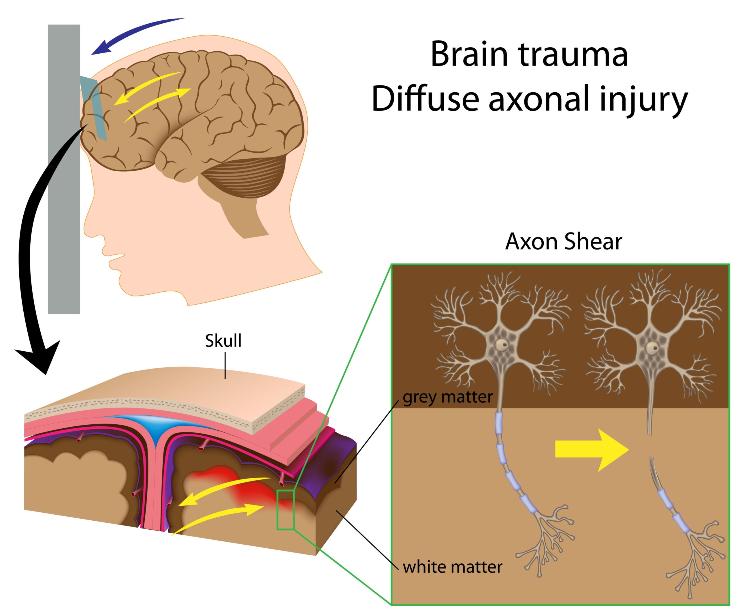
The pathophysiology of TBI is complex. Initially, the brain can suffer from direct mechanical injury, which results in cellular disruption, inflammation, and neuronal death. Following the initial trauma, secondary injury processes can exacerbate the damage, leading to further neuronal injury due to factors such as metabolic disturbances and elevated intracranial pressure. Identifying the precise location and severity of these injuries is crucial for effective treatment and rehabilitation strategies, as TBI can lead to long-term neurological deficits and a diminished quality of life.
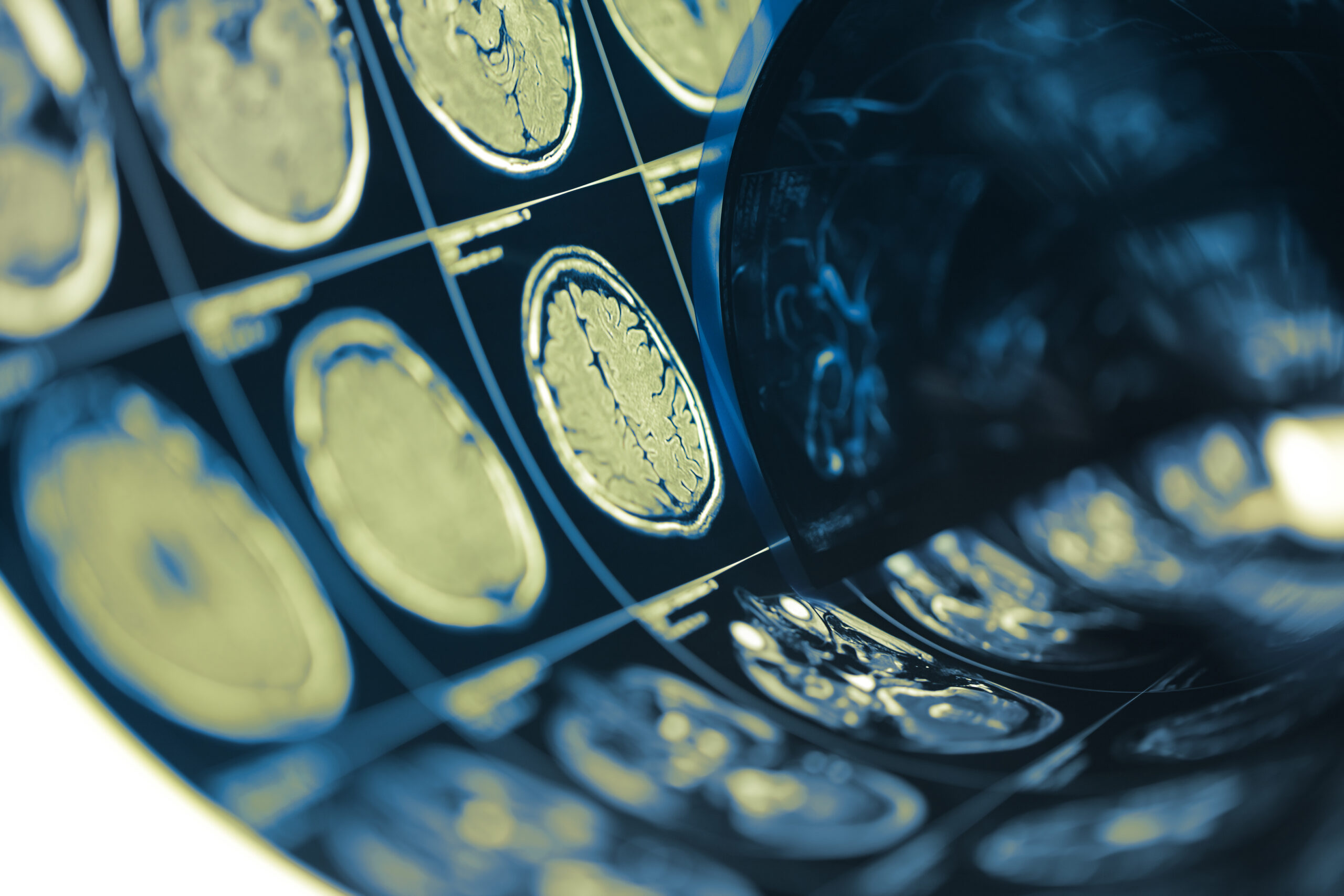
Diagnostic tools for TBI have historically relied on imaging techniques such as computed tomography (CT) and magnetic resonance imaging (MRI). While CT is effective for detecting acute hemorrhages and certain structural injuries, it may not adequately visualize subtle changes or diffuse axonal injury. MRI provides detailed imaging of the brain’s soft tissues and can identify lesions that CT might miss. However, both techniques have limitations in terms of accessibility, speed, and the ability to provide real-time monitoring of injury evolution.
Recent advancements in neuroimaging are exploring innovative methods to enhance the detection and characterization of TBI. Among these, Magnetic Particle Imaging (MPI) emerges as a promising approach. MPI utilizes superparamagnetic nanoparticles for imaging, allowing for real-time, high-resolution visualization of brain injury sites. This could herald a new era in TBI diagnostics, offering insights not only into the injury itself but also into the underlying biological processes, thus paving the way for targeted therapeutic interventions.
Magnetic Particle Imaging Technique
Magnetic Particle Imaging (MPI) is an innovative imaging technique that leverages the unique properties of superparamagnetic nanoparticles to achieve high-resolution, real-time imaging of biological tissues. This technology stands out due to its ability to provide visualization of tissues with minimal background noise and significant depth penetration, making it particularly advantageous for brain imaging applications. At the core of MPI is the use of these nanoparticles, which can be precisely targeted and controlled through magnetic fields. When these particles are introduced to the body, they accumulate in areas of interest, such as those affected by traumatic brain injury (TBI).
One of the key elements of MPI is its robust safety profile compared to traditional imaging modalities. Unlike methods that utilize ionizing radiation, such as computed tomography (CT), MPI employs low magnetic fields that pose no risk of radiation exposure. This aspect is especially relevant when considering populations such as children or individuals requiring repeated imaging due to ongoing health issues.
The MPI imaging process begins with the injection of superparamagnetic nanoparticles into the bloodstream. As these particles circulate, they preferentially accumulate in injured or damaged tissues within the brain. The MPI system generates magnetic fields to localize and detect the particles, subsequently creating detailed images based on their distribution and concentration. The resulting images reveal not only the location of the injury but also its extent and potential involvement of surrounding tissues, providing a comprehensive view of the traumatic impact.
Another significant advantage of MPI is its rapid imaging capabilities. Traditional imaging methods often require significant preparation time and may involve delays in obtaining essential diagnostic information. In contrast, MPI can deliver real-time results, enabling clinicians to make prompt decisions that can drastically alter patient outcomes, especially in acute settings where time is of the essence.
The resolution achieved by MPI is another noteworthy feature. It allows for the visualization of structures and lesions at a microscopic level, which is critical for detecting minor injuries, such as diffuse axonal injury, that are often missed by CT or even some MRI techniques. This meticulous detail enhances the understanding of cellular responses to TBI, which could lead to better-targeted therapies based on the specific nature of the injury.
In addition to its clinical implications, MPI has potential applications in understanding the biological processes associated with brain injuries. By monitoring how the nanoparticles interact with various cellular structures over time, researchers can gain insights into the dynamics of repair mechanisms and inflammatory responses following TBI. This could foster the development of new therapeutic strategies aimed at mitigating the destructive cascade following brain injuries.
As the technology continues to evolve, ongoing research aims to enhance the functionality of MPI systems, potentially integrating them with other imaging modalities or therapeutic applications. Such advancements could pave the way for comprehensive systems that not only visualize injuries in real-time but also deliver targeted treatment, transforming the landscape of TBI management.
Results and Analysis
In this study, the application of Magnetic Particle Imaging (MPI) to detect in vivo traumatic brain injury foci yielded promising results that could redefine diagnostic protocols for TBI. The MPI technique demonstrated the ability to resolve nuances in brain injuries that traditional imaging modalities often overlook. The selection of superparamagnetic nanoparticles proved effective, with these particles showing a high affinity for the damaged tissues, thus enabling precise localization of injury sites.
Following the administration of the nanoparticles, a series of MPI scans revealed significant areas of accumulation corresponding to sites of traumatic injury, particularly in regions commonly associated with TBI, such as the frontal and temporal lobes. The images produced displayed both the extent and severity of the lesions with unprecedented clarity, as compared to MRI or CT scans that exhibited limitations in such subtle visualizations. This capability underscores MPI’s potential as a more sensitive imaging modality capable of detecting even minor injuries that can have profound clinical implications.
The analysis further indicated a correlation between the density of nanoparticle accumulation and the severity of the traumatic injury, observed through a quantifiable increase in the magnetic signal in areas of higher damage. This relationship suggests that MPI not only provides qualitative images but can also facilitate a quantitative assessment of injury severity, which is a vital component in guiding treatment decisions. Such quantifiable data may enhance clinical decision-making by allowing for personalized treatment approaches tailored to the specific injury profile of each patient.
Additionally, MPI’s real-time imaging capability significantly reduced the time required to acquire critical information compared to traditional imaging techniques. In acute settings, where rapid diagnosis is crucial, MPI allowed for immediate assessment of the injury location and severity, providing clinicians with the data needed to initiate timely interventions. The speed of MPI was further evidenced in cases where acute intracranial hemorrhage or significant swelling presented, enabling quick evaluations of treatment responses.
Another critical aspect analyzed was the safety and biocompatibility of the superparamagnetic nanoparticles. Throughout the study, no adverse reactions were documented post-injection, indicating that the nanoparticles are safe for use in clinical settings. This finding aligns with research suggesting that such nanoparticles can be safely utilized without the risks associated with ionizing radiation that accompany CT imaging.
The results not only demonstrate MPI’s potential as a robust imaging tool for TBI but also suggest its applicability in exploring associated biological processes following brain injury. The tracking of real-time interactions between the nanoparticles and cellular environments may yield insights into ongoing neuroinflammatory responses, thus contributing to a deeper understanding of the pathophysiology of TBI.
Future studies will be essential to further validate these findings on a larger cohort and to explore the long-term implications of MPI findings in clinical practice. Continued exploration into the integration of MPI with therapeutic modalities could lead to groundbreaking advances in how TBI is diagnosed and treated.
Future Directions and Applications
The landscape of traumatic brain injury (TBI) management is on the brink of transformation, with Magnetic Particle Imaging (MPI) paving the way for novel applications that extend beyond mere diagnostics. As research continues to build upon the foundation established by earlier studies, several promising avenues emerge, particularly in personalized medicine, therapeutic monitoring, and enhanced multimodal imaging strategies.
One of the foremost applications of MPI lies in its potential to inform personalized treatment protocols for TBI. The ability to quantitatively assess the severity and specific characteristics of brain injuries offers clinicians a refined tool that can significantly enhance decision-making. By tailoring interventions based on real-time MPI findings, clinicians can potentially optimize treatment plans, adjusting therapies to suit the individual needs of patients based on their unique injury profiles. This personalized approach could lead to more effective rehabilitation strategies and improved long-term outcomes for patients suffering from TBI.
Moreover, MPI’s role in therapeutic monitoring is an area ripe for exploration. The technology’s capacity to visualize changes in the brain over time opens up opportunities for monitoring the efficacy of various medications and interventions aimed at promoting recovery post-injury. For instance, as researchers develop new neuroprotective agents or therapies targeting inflammatory responses in TBI, MPI could serve as a real-time assessment tool, allowing for the observation of treatment effects and adaptive modifications in therapeutic approaches. Such capabilities could greatly enhance clinical trials, providing immediate feedback on how specific treatments influence injury healing and recovery trajectories.
Integrating MPI with existing imaging modalities further exemplifies another direction for future research. The combination of MPI with techniques like MRI or positron emission tomography (PET) could yield comprehensive insights into both the structural and functional aspects of brain injuries. Such integration would enable a more holistic perspective, elucidating not only where damage occurs but also how it affects brain function and connectivity. This synergy could enhance diagnostic accuracy and lead to refined therapeutic interventions aligned with the neuroanatomical and physiological context of each patient’s injury.
Additionally, MPI could play a pivotal role in neuroscience research by facilitating investigations into the underlying mechanisms of TBI and subsequent recovery. The real-time visualization of how superparamagnetic nanoparticles interact with damaged neural tissue could shed light on critical processes such as neuroinflammation, cellular repair, and neuronal regeneration. Insights garnered from such studies could contribute to the development of innovative therapeutic strategies focused on enhancing recovery, thereby addressing some of the long-term disabilities associated with TBI.
As MPI technology continues to advance, considerations around its scalability and accessibility will be important. Future efforts should focus on refining the MPI equipment to ensure it can be implemented in a variety of clinical settings, including emergency departments and remote medical facilities. Enhancing the technological infrastructure associated with MPI could bridge the gap in TBI management, particularly in underserved regions where sophisticated imaging may be limited.
The future of MPI in the context of traumatic brain injury is filled with potential. As researchers and clinicians harness its capabilities, the integration of this cutting-edge imaging modality into clinical practice could revolutionize how TBI is diagnosed, monitored, and treated, ultimately leading to better patient outcomes and a deeper understanding of brain injury pathophysiology.