Study Overview
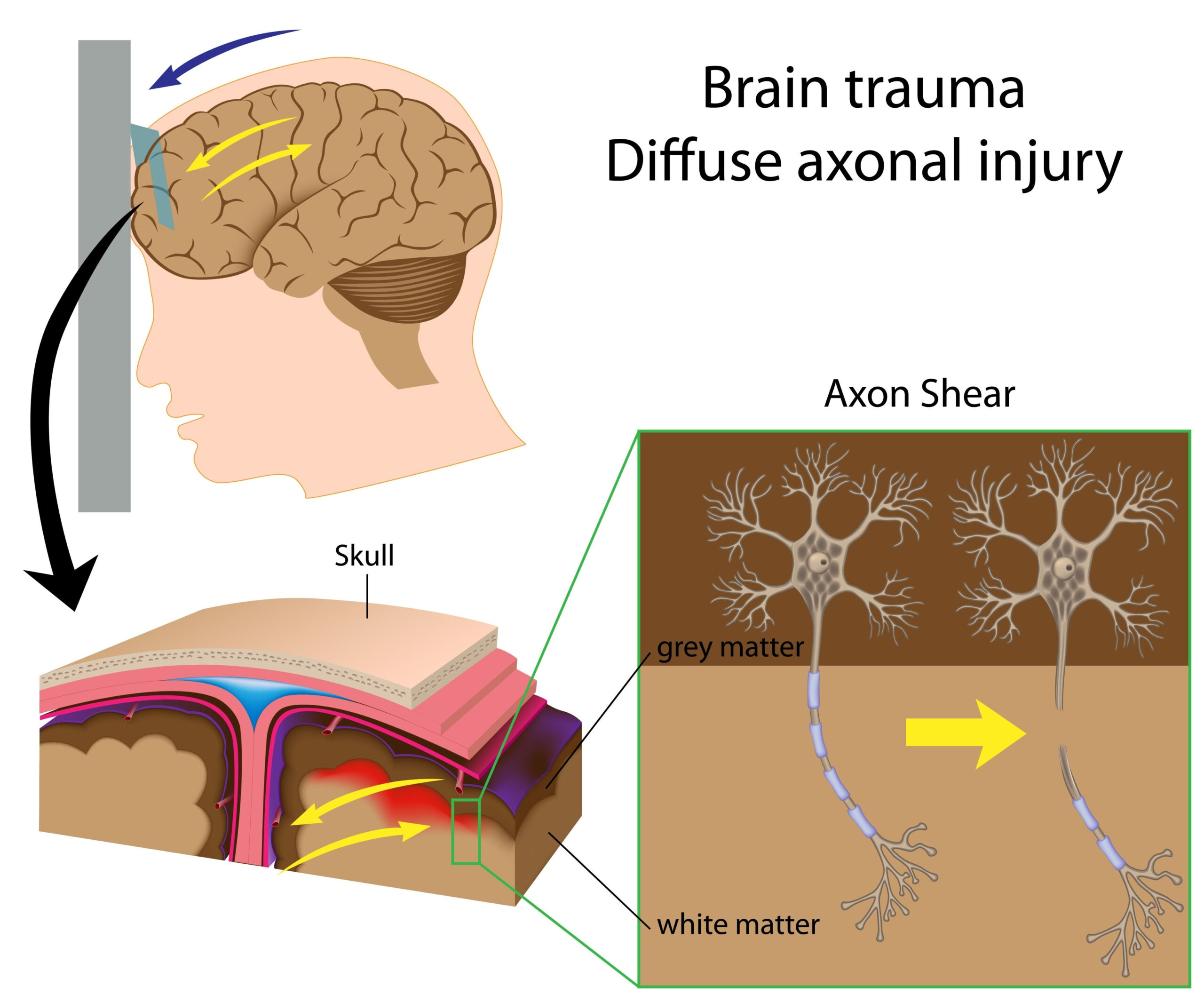
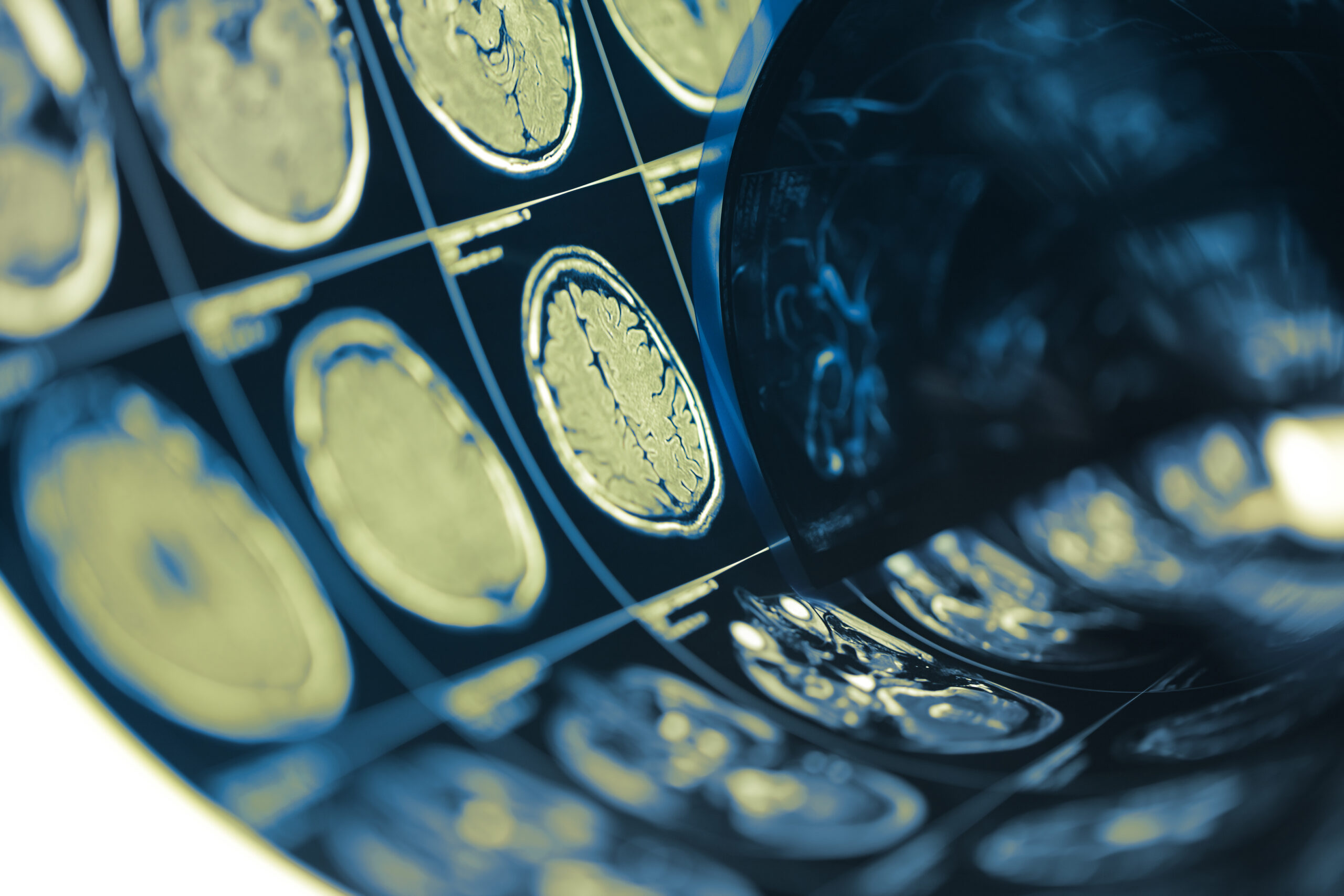
This study investigates the integration of dynamic brain functional imaging and electroencephalography (EEG) spectral parameters as a novel approach for identifying early-stage brain injuries. The research aims to enhance diagnostic accuracy and provide valuable insights into pathological changes that occur in the brain following trauma. Traditional methods for assessing brain injuries often rely on imaging techniques such as MRI or CT scans, which, while effective in revealing structural abnormalities, may not adequately capture the functional changes occurring at a microlevel. This study addresses this gap by combining insights from both brain imaging and EEG, which measures the electrical activity of the brain.
By focusing on dynamic imaging methodologies, the researchers highlight how brain activity and connectivity patterns can fluctuate over time, offering a more comprehensive understanding of brain function immediately following injury. The combination of functional imaging techniques with EEG spectral analysis aims not only to detect the presence of injury but also to evaluate the severity and potential impact on cognitive functions. Through an extensive analysis of existing data alongside new experiments, the study seeks to validate the efficacy of this integrated approach, laying the groundwork for improved screening and monitoring protocols in clinical settings.
Ultimately, this research endeavors to pave the way for more efficient tools that can be employed in emergency and rehabilitation settings, potentially transforming how clinicians assess and manage brain injuries in patients. By emphasizing early detection and ongoing monitoring, the findings may contribute to better outcomes in patient care and highlight the importance of both functional and structural assessments in understanding brain health.
Methodology
The study employs a robust methodology focusing on the integration of advanced imaging techniques and EEG spectral analysis. Initially, participants diagnosed or suspected of having sustained a brain injury underwent both functional imaging and EEG assessments. Functional imaging, utilizing either positron emission tomography (PET) or functional magnetic resonance imaging (fMRI), captured real-time brain activity and blood flow dynamics, providing critical insights into how various brain regions communicate and perform interconnected tasks.
Simultaneously, EEG was used to record electrical activity from the scalp, allowing researchers to measure brain wave patterns across different frequency bands, such as delta, theta, alpha, beta, and gamma. These spectral parameters are crucial as they reflect the brain’s response to injury and can reveal alterations associated with cognitive impairment and emotional disturbances following trauma. By combining imaging data with EEG results, researchers aimed for a multifaceted understanding of the brain’s functional state.
Participants were subjected to a series of controlled tasks designed to provoke specific cognitive and emotional responses. This was essential to observe how brain functionality varied in real-time, especially under stressors similar to what might occur following an injury. Dynamic functional connectivity was assessed through the analysis of temporal patterns in brain networks, allowing researchers to identify abnormal connectivity profiles that might correlate with injury severity.
To ensure the findings were reliable and valid, statistical analyses were performed, including correlations between EEG spectral changes and functional imaging outcomes. The sample size was determined based on power calculations to ensure that the results could be generalized to a broader population. Participants were also categorized based on injury severity, which provided a nuanced analysis of how different levels of brain impairment manifested in terms of both structural and functional changes.
Moreover, the study incorporated longitudinal assessments. Follow-up evaluations were conducted at set intervals post-injury to monitor changes in brain function and to assess the recovery trajectory. This approach enabled the researchers to investigate the time-dependent nature of brain healing and recovery, providing critical insights into the temporal dynamics of brain activity changes following an injury.
Ethical considerations were paramount throughout the study. Informed consent was obtained from all participants, and the research protocol was reviewed and approved by an institutional ethics board. Safety measures were implemented to minimize any potential risks associated with both imaging techniques and EEG recordings, ensuring the well-being of participants during the study.
The methodology used in this research highlights its innovative approach to combining advanced imaging and EEG analysis, thus potentially advancing the paradigm of early diagnosis and monitoring of brain injuries. This integrated approach stands to fundamentally alter conventional practices, emphasizing the necessity for both functional assessments and traditional imaging in the full evaluation of brain health.
Key Findings
The research yielded several significant findings that bolster the hypothesis that combining dynamic brain functional imaging with EEG spectral analysis can enhance the detection and understanding of early-stage brain injury. One of the primary revelations was the identification of significant variations in functional connectivity patterns among participants with different severity levels of brain injuries. Specifically, individuals with more pronounced injuries exhibited disrupted connectivity within critical brain networks, particularly those associated with cognitive functions such as attention and memory. These disruptions were quantified using advanced analytical techniques that evaluate how different brain regions communicate, providing a clearer picture of the brain’s functional state post-injury.
Furthermore, the analysis of EEG spectral parameters showed notable changes correlating with the degree of injury. In patients with mild traumatic brain injuries, increases in beta and gamma wave activity were observed, which suggest heightened cortical excitability and may indicate an attempt at compensatory cognitive processing. Conversely, a reduction in alpha rhythm, commonly related to relaxation and inhibition of irrelevant stimuli, was documented, indicating potential difficulties in cognitive control and focus among those with more severe injuries. These spectral changes are critical, as they align with current theories regarding the brain’s adaptive versus maladaptive responses following trauma.
The longitudinal assessment provided insights into the recovery trajectories of participants. Notably, those who showed a normalization of functional connectivity and EEG patterns over time tended to report better cognitive and emotional outcomes. Such findings underscore the relevance of ongoing monitoring as a tool for predicting recovery, as well as the potential benefits of early intervention strategies tailored to individual recovery profiles. In contrast, participants who maintained abnormal connectivity patterns displayed more persistent cognitive impairments, emphasizing the need for targeted therapeutic approaches that address dysfunctional brain activity.
In addition to connectivity and spectral changes, the integrated approach revealed patterns that transcended traditional diagnostic classifications. For instance, certain EEG profiles were found to be predictive of later cognitive deficits even when structural imaging appeared normal, showcasing a critical advantage of integrating functional assessments into clinical workflows. This capability to detect underlying dysfunction that might be missed by standard imaging techniques is a significant leap forward in the timely and effective management of brain injuries.
Collectively, these findings suggest that the integration of dynamic functional imaging with EEG spectrography not only enhances diagnostic accuracy but also provides profound insights into the mechanisms driving brain injuries. This holds implications for future research to further investigate specific interventions aimed at improving functional outcomes for patients experiencing early-stage brain injuries. The promise of personalized treatment plans tailored to the unique brain activity patterns of individuals represents a significant advancement in the field of neurorehabilitation.
Clinical Implications
The integration of dynamic brain functional imaging and EEG spectral parameters offers numerous clinical implications that can significantly impact the evaluation and management of brain injuries. Primarily, this dual approach enhances the diagnostic toolkit available to clinicians, allowing for earlier and more accurate identification of brain injuries that might otherwise go unnoticed when using standard imaging techniques alone. The ability to capture real-time fluctuations in brain connectivity and activity promotes a thorough understanding of how injuries affect individual patients, thereby facilitating tailored treatment strategies that cater to specific needs based on their unique brain profiles.
Moreover, the data gleaned from this combined methodology can serve as a critical predictor for recovery trajectories. By establishing baseline functional and spectral parameters shortly after a brain injury, clinicians can monitor changes over time and correlate these with patient outcomes. This longitudinal aspect allows healthcare providers to adjust rehabilitation plans dynamically, optimizing interventions as more information becomes available about each patient’s response to treatment. For instance, if ongoing EEG assessments demonstrate habitual patterns of abnormal connectivity associated with cognitive deficits, clinicians may prioritize specific cognitive therapies or behavioral interventions to counteract these issues.
Furthermore, the opportunity to differentiate between adaptive and maladaptive responses to injury can inform treatment approaches. Understanding which patients are exhibiting increased cortical excitability, for example, can prompt clinicians to consider strategies that capitalize on this heightened activity, potentially harnessing it to promote recovery. Conversely, identifying patients who display reduced brain wave activity in critical areas could lead to early intervention programs aimed at bolstering cognitive functions, including attention and memory exercises designed to reactivate these regions.
This innovative methodology does not just assist in immediate diagnosis and treatment; it can also influence the discharge planning and long-term care pathways for patients with brain injuries. The continuous assessment of functional and spectral changes post-injury can inform decisions regarding return-to-work protocols, educational accommodations, and even the need for psychological support, thereby creating a more nuanced and comprehensive care plan that responds to the patient’s ongoing needs.
Importantly, the integration of functional imaging with EEG brings forth the potential to develop standardized screening tools that can be disseminated across clinical settings. These tools can establish benchmarks for brain health and functionality, serving as critical reference points for clinicians worldwide. As more healthcare facilities adopt these advanced techniques, the normalization of such assessments in routine clinical practice could lead to a paradigm shift in how brain injuries are understood, treated, and monitored over time, ultimately fostering better patient outcomes and quality of life.
The clinical implications of utilizing this integrated approach are profound, paving the way for more precise, individualized, and effective treatment pathways for patients with brain injuries, and supporting a proactive stance towards neurorehabilitation that prioritizes functional recovery and cognitive health.