Study Overview
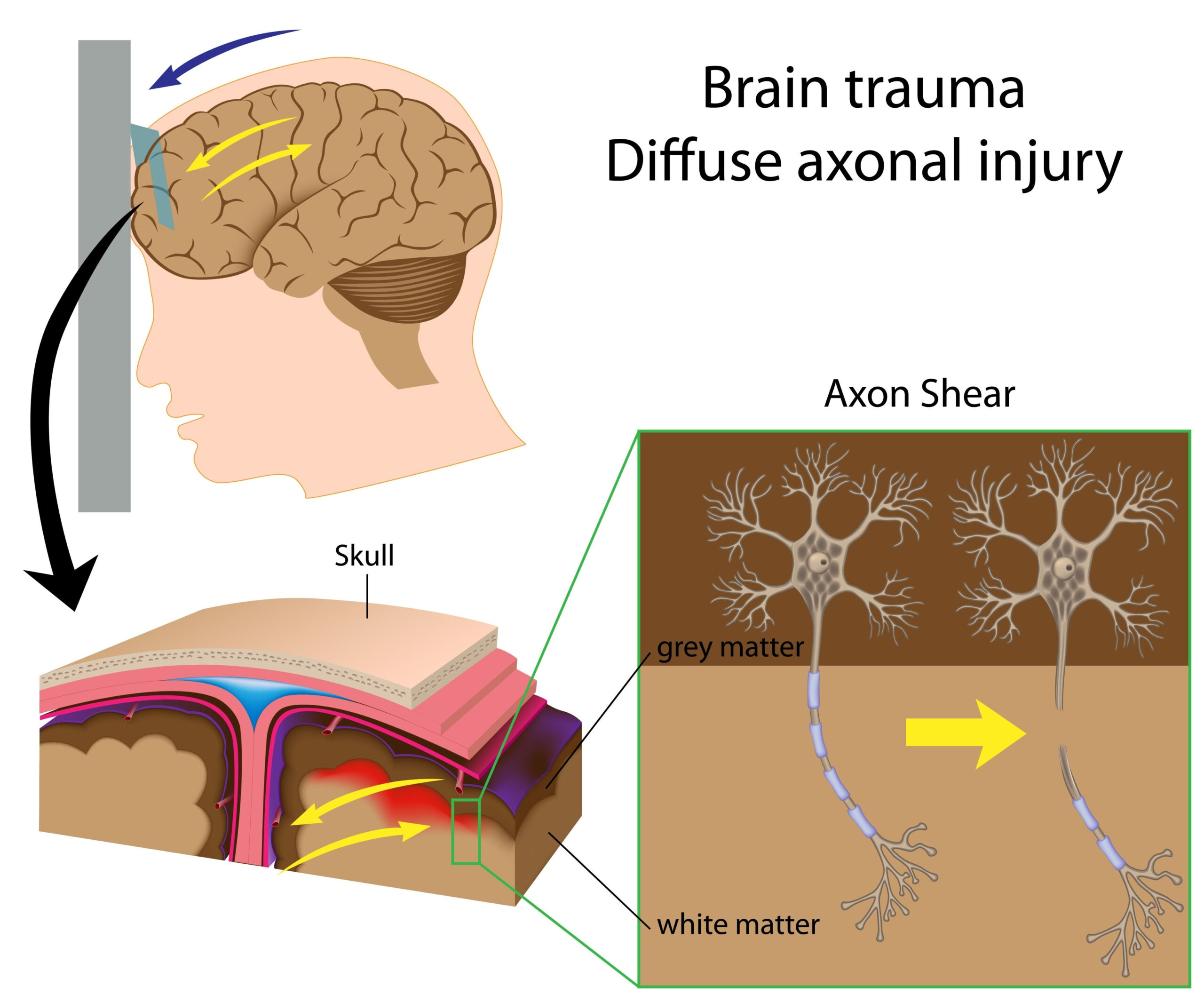
The research investigates the complex biological processes that occur in human astrocytes following traumatic brain injury (TBI). Specifically, it focuses on the delayed neurotoxic effects mediated by lipids, which are crucial components of cell membranes and play significant roles in signaling pathways. The study utilizes an innovative “in-a-dish” model to simulate TBI, allowing for detailed observation of astrocyte behavior in a controlled environment that mimics in vivo conditions. This model demonstrates how injury to astrocytes can lead to a cascade of lipid-related neurotoxic events over time, potentially worsening brain damage long after the initial insult. The findings aim to contribute to a deeper understanding of the temporal dynamics of neurotoxicity in brain injuries, with an emphasis on identifying processes that may be targeted for therapeutic intervention. Thus, the study highlights the importance of delving into the mechanisms of lipid-mediated neurotoxicity as a significant factor in post-injury degeneration of brain tissue, paving the way for future research and treatment strategies aimed at improving outcomes for patients with TBI.
Methodology
The investigation employed a cutting-edge in vitro model designed to replicate the complex environment surrounding astrocytes during traumatic brain injury. Human astrocytes, cultured from brain tissue samples, were utilized to ensure that the findings were relevant to human physiology. The model employed a mechanical stimulus mimicking the physical impact associated with TBI, allowing for the assessment of immediate cellular responses as well as the progression of delayed neurotoxic effects over time.
Astrocytes were subjected to this mechanical insult, after which samples were harvested at predetermined intervals to examine the resultant biochemical changes. To understand the lipid-mediated toxicity mechanisms, lipid profiles were analyzed using advanced mass spectrometry techniques. This allowed for the identification of specific lipid species that were either elevated or depleted following injury, making it possible to assess their correlation with neurotoxic effects. Additionally, various assays were conducted to measure markers of cell viability, apoptosis, and inflammation, providing a comprehensive assessment of astrocyte health and function in response to injury.
Fluorescent imaging techniques were implemented to visualize astrocytic changes at the cellular level, such as alterations in morphology and activation states. These imaging studies complemented the biochemical analyses, revealing how injury transformed astrocyte behavior over time—specifically how they transitioned from a protective to a detrimental role in the context of neuroinflammation and neurodegeneration.
Statistical analyses were performed to determine the significance of observed changes, ensuring that results were robust and reliable. Different experimental groups were established to differentiate between immediate responses to injury and those that occurred after a delay, reinforcing the hypothesis that lipid-mediated neurotoxicity plays a crucial role in the pathophysiological processes following TBI. The combined approach of mechanical injury, lipid profiling, molecular assessments, and imaging analysis furnished a holistic view of the injury dynamics affecting human astrocytes, laying the groundwork for further explorations into targeted therapeutic strategies.
Key Findings
The investigation yielded several noteworthy findings that deepen our understanding of the neurotoxic processes initiated by traumatic brain injury (TBI) in human astrocytes. Central to these findings is the observation that lipid mediators become significantly altered following mechanical injury to astrocytes. Specifically, certain lipid species, such as ceramides and sphingomyelins, were found to increase markedly post-injury. These lipids are not merely structural components; they act as signaling molecules that can contribute to cellular stress and inflammation, implicating them in the progressive degeneration observed after TBI.
Further analyses revealed the timeline of these lipid changes, demonstrating that alterations were not confined to the immediate aftermath of injury. For instance, while some lipids exhibited acute fluctuations, others displayed a delayed increase that correlated with heightened inflammatory responses and apoptosis markers. One specific finding indicated that astrocytes shifted from a neuroprotective role to a state of neurotoxicity, exacerbated by an accumulation of pro-inflammatory cytokines. This shift underscores the duality of astrocyte function: while they are crucial for maintaining neuronal health, their response to injury can become maladaptive, perpetuating damage rather than facilitating repair.
Additionally, the research highlighted the significant relationship between lipid alterations and markers of cell viability. Decreases in specific phospholipids were strongly associated with reduced astrocytic survival, indicating that lipid composition is directly linked to cell health following traumatic injury. These correlations were substantiated through statistical analyses, showing that the degree of lipid alteration can serve as a predictive factor for the extent of neurodegeneration observed in the cells.
Fluorescent imaging further illuminated these dynamic changes at a cellular level. The imaging studies depicted a progressive morphological transformation of astrocytes from a star-shaped, healthy state to a more reactive form characterized by cell swelling and increased branching. This morphological shift was paralleled by a rise in the expression of glial fibrillary acidic protein (GFAP), a marker commonly associated with astrocyte activation. Notably, the degree of GFAP expression also reflected the extent of lipid accumulation, suggesting that monitoring lipid profiles could provide insights into astrocyte activation states.
In summary, the study delineates a complex interplay between lipid metabolism, astrocytic responses, and the resulting neurotoxic environment following TBI. The findings suggest that clinically, there may be potential for targeting lipid pathways to mitigate secondary damage in TBI patients. Future research will be essential to translate these findings into therapeutic strategies, potentially enabling interventions that can modulate astrocyte function and mitigate delayed neurotoxicity in the aftermath of brain injuries.
Clinical Implications
The findings from this research have significant clinical implications, particularly in the context of developing targeted therapies for traumatic brain injury (TBI) management. The identification of delayed lipid-mediated neurotoxicity highlights a critical window of intervention opportunities that may have previously been overlooked in treating TBI. Understanding the role of specific lipid mediators, such as ceramides and sphingomyelins, in the neurotoxicity cascade not only enhances our comprehension of the pathophysiology of TBI but also paves the way for novel therapeutic strategies aimed at mitigating secondary brain damage.
Clinical interventions could focus on modulating lipid profiles in astrocytes to promote neuroprotection over neurotoxicity. For instance, pharmacological agents that can inhibit the synthesis of harmful lipid species or restore levels of protective phospholipids may hold promise in preserving astrocyte function. This approach could be particularly beneficial during the critical phases following mechanical injury when the shift from protective to harmful astrocytic roles occurs. By targeting lipid metabolism pathways, it may be possible to alter the trajectory of neuroinflammation and neuronal degeneration, thereby improving patient outcomes.
Furthermore, the research underscores the importance of early biomarker identification for assessing the extent of astrocytic damage and neurodegeneration following TBI. Monitoring lipid alterations in the cerebrospinal fluid or serum could provide clinicians with valuable insights into the timing and severity of injury, allowing for more informed decisions regarding treatment interventions. Establishing lipid profiles as biomarkers may facilitate a tailored approach to therapy, where interventions can be personalized based on individual lipid signatures and the corresponding neurotoxic risk.
Additionally, as the study reveals a profound relationship between astrocyte morphology, activation markers like GFAP, and lipid accumulation, there is potential for utilizing imaging techniques in clinical settings. Advanced imaging modalities could be developed or adapted to visualize and quantify lipid changes and astrocytic activation in real-time, offering a non-invasive method to monitor the effectiveness of treatments aimed at reversing or preventing astrocytic degeneration.
In summary, the exploration of lipid-mediated neurotoxicity in astrocytes offers new avenues for therapeutic intervention in TBI management. By targeting lipid pathways, enhancing biomarker identification, and employing advanced imaging techniques, the medical field can move towards more effective strategies to combat the complications associated with traumatic brain injuries. This knowledge could ultimately lead to significant advancements in patient care, improving recovery trajectories for individuals affected by TBIs.