Anatomy of the Second Cranial Nerve
The second cranial nerve, also known as the optic nerve, is a pivotal component of the visual system. It is not a true cranial nerve but rather an extension of the brain parenchyma, which transmits electrical impulses from the retina to the brain to be processed into visual information[3].
Intraocular and Intracranial Pathway
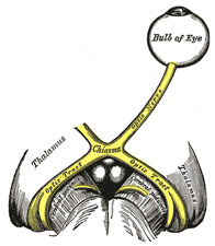
The optic nerve consists of axons of retinal ganglion cells (RGCs) that extend from the RGC soma to the lateral geniculate nucleus (LGN), where the neurons synapse. The nerve is subdivided into fascicles by connective tissue and glial septa and is surrounded by cerebrospinal fluid (CSF). The optic nerve has several segments: intraocular, intraorbital, intracanalicular, and intracranial, with a total length of approximately 50 mm[7].
Blood Supply
The blood supply to the optic nerve includes the central retinal artery, the Zinn-Haller circle formed by the short posterior ciliary artery extensions, and pial vessels[7].
Visual Pathway
The visual pathway begins with the optic nerve, which captures visual stimuli from the surroundings. These stimuli are processed by a system of interconnecting neurons, starting from the optic nerve and culminating at the visual processing center in the forebrain known as the visual cortex[12].
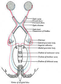
After the optic nerve exits the eye, it passes through the optic canal to reach the optic chiasm, where fibers from the nasal halves of each retina cross to the opposite side. The fibers then continue as the optic tracts, which wrap around the midbrain to reach the LGN. From the LGN, the optic radiations fan out to the primary visual cortex in the occipital lobe[12].
Functional Anatomy and Neurosurgical Approaches
The optic nerve’s functional anatomy is critical for neurosurgical interventions. Surgical manipulation of the visual pathway requires detailed knowledge of the nerve’s anatomy to avoid iatrogenic injuries. This is particularly important when treating major lesions affecting the optic nerve, such as tumors, intracranial hypertension, trauma, and aneurysms[5].
Clinical Relevance and Imaging
MRI plays a crucial role in diagnosing lesions in the optic nerves, as the clinical definition of a lesion is often not clear. The optic nerves can be affected by various diseases, including congenital malformations, inflammatory, vascular, and neoplastic diseases[3].
Binocular Integration and Visual Field Representation
The visual pathway is also involved in binocular integration, which is the process of combining visual information from both eyes to create a single, cohesive visual perception. Despite severe lesions in the geniculostriate pathway, such as in the case of patient IB with left eye microphthalmia, the primary visual cortex can still maintain a complete visual field representation, indicating the flexibility of developing retinotopic maps[6].
Direct Communication with the Subarachnoid Space
There is evidence of direct communication between the CSF of the subarachnoid space and the extravascular space of the human visual pathway. This communication is essential for the functioning of the glymphatic system, which is involved in the clearance of metabolic waste from the central nervous system[8].
In summary, the second cranial nerve is a complex structure that plays a vital role in the visual system, from capturing visual stimuli to processing them into images in the brain. Its intricate anatomy and the pathways it forms are essential for vision and are of significant interest in clinical and surgical contexts.
Citations:
[1] https://www.semanticscholar.org/paper/ca807bcb42a9fb028112ba407c7920fbc04d473c
[2] https://www.semanticscholar.org/paper/cb842e55f4058b5b7aae1395989115d40cb4a067
[3] https://pubmed.ncbi.nlm.nih.gov/36116850/
[4] https://pubmed.ncbi.nlm.nih.gov/38194453/
[5] https://www.semanticscholar.org/paper/eb1635303558984642113984f397a0d1f362f50a
[6] https://www.ncbi.nlm.nih.gov/pmc/articles/PMC8835673/
[7] https://www.semanticscholar.org/paper/9cc32e5faa48cecda3930afd467053ef82f8a270
[8] https://pubmed.ncbi.nlm.nih.gov/31247084/
[9] https://www.semanticscholar.org/paper/c9637ea87bdb2fb7facac4052f3be4e3640d0059
[10] https://pubmed.ncbi.nlm.nih.gov/32367252/
[11] https://www.semanticscholar.org/paper/dd75dace2b0fa1ac908778d296e6dc71e9cb413e
[12] https://www.semanticscholar.org/paper/5e1023639bb432609a14ad71dbd7eeaa71fbcd85
[13] https://www.semanticscholar.org/paper/c1bfb283ab9f944d24adcf7032a55ea21b0591e8
[14] https://www.ncbi.nlm.nih.gov/pmc/articles/PMC8448312/
[15] https://pubmed.ncbi.nlm.nih.gov/24678025/
[16] https://pubmed.ncbi.nlm.nih.gov/31427779/