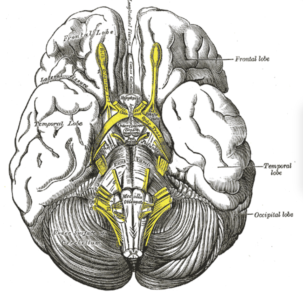
Detailed Analysis of the Anatomy and Course of the Olfactory Nerve
The olfactory nerve, or Cranial Nerve I, is unique among cranial nerves as it is solely sensory and dedicated to the sense of smell. This nerve originates from the olfactory epithelium in the nasal cavity. The olfactory epithelium contains olfactory receptor cells, which are neurons equipped with odorant receptors. These cells extend their axons through the cribriform plate of the ethmoid bone to reach the olfactory bulb, which lies at the base of the brain directly above the nasal cavity[1][7].
The olfactory bulb is a crucial structure where the first synaptic relay of olfactory information occurs. From the olfactory bulb, nerve fibers, known as the olfactory tract, project to various parts of the brain, including the primary olfactory cortex, which is responsible for the perception and identification of odors. This direct pathway to the brain allows for the immediate processing of olfactory information, which is essential for survival functions such as detecting food and environmental dangers[1][7].
Course of the Olfactory Nerve
The course of the olfactory nerve begins at the olfactory mucosa, where olfactory receptor neurons detect odorant molecules. These neurons send their axons through the cribriform plate, a sieve-like structure that forms part of the ethmoid bone. The axons bundle together to form the fila olfactoria, which are small nerve fiber bundles that collectively constitute the olfactory nerve[1][7].
Upon passing through the cribriform plate, the axons enter the olfactory bulb. Here, they synapse with second-order neurons known as mitral and tufted cells. This synaptic interaction occurs within structures called glomeruli, which are spherical units where information about specific odors is processed[1][7].
From the olfactory bulb, the olfactory tract extends posteriorly along the base of the frontal lobe to reach the primary olfactory cortex. This tract also sends offshoots to other limbic areas, including the amygdala and entorhinal cortex, which are involved in emotional and memory-related aspects of olfactory processing[1][7].
Microsurgical Relevance
The olfactory nerves are vulnerable to damage due to their location and the thinness of the cribriform plate. Surgical approaches to the anterior cranial fossa, particularly for the removal of tumors or during nasal surgeries, require careful manipulation to avoid damaging these nerves. Understanding the detailed anatomy of the olfactory nerve and its protective coverings, such as the outer arachnoid envelope, is crucial for surgeons. This knowledge helps in planning surgical approaches that minimize harm to olfactory function, which can significantly affect a patient’s quality of life if impaired[1][17].
In summary, the olfactory nerve plays a critical role in the sensory system by detecting odors and transmitting this information to the brain for further processing. Its unique anatomy and course through the cribriform plate to the olfactory bulb and then to various brain regions highlight its importance in olfactory perception and its vulnerability to surgical interventions.
Sources
[1] The Olfactory Nerve: Anatomy and Pathology. https://pubmed.ncbi.nlm.nih.gov/36116849/
[2] Spontaneous r ecovery of Medial Prefrontal Syndrome f ollowing Giant Olfactory Groove Meningioma r esection : A c ase r eport https://www.semanticscholar.org/paper/402d793500655abba39f605172f8d41d7f1c6946
[3] Brief sensory deprivation triggers cell type-specific 2 structural and functional plasticity in olfactory bulb neurons Neuromodulation of Spike-Timing-Dependent Relocation axon initial segment structure and function by multiplexed Monophasic dopaminergic cells (putative anaxonic) Biphasic dopaminerg https://www.semanticscholar.org/paper/a23c30f4e632c43a33f98cedaed4948776ad5573
[4] Olfactory nerve schwannoma: how human anatomy and electron microscopy can help to solve an intriguing scientific puzzle https://pubmed.ncbi.nlm.nih.gov/35913116/
[5] Olfactory Nerves (I) https://www.semanticscholar.org/paper/5a92d1c14735e3feecd926b94ba47bf96fe3ed0d
[6] Ablation of Mouse Adult Neurogenesis Alters Olfactory Bulb Structure and Olfactory Fear Conditioning https://www.ncbi.nlm.nih.gov/pmc/articles/PMC2858604/
[7] Anatomy of the olfactory nerve: A comprehensive review with cadaveric dissection https://pubmed.ncbi.nlm.nih.gov/29088516/
[8] [Morphological studies on the distribution of the olfactory nerves in Japanese adults and fetuses]. https://pubmed.ncbi.nlm.nih.gov/3998082/
[9] The structure and function of neural connectomes are shaped by a small number of design principles https://www.semanticscholar.org/paper/8663ee4d60f3ec34a39b8ec56d9efc5545d66b21
[10] Overview of Anatomy and Physiology of Gustatory and Olfactory System https://www.semanticscholar.org/paper/0b56d61bca3e01dd2700ebb1a2ea793d65f865e7
[11] New Antiseptic Artificial Membrana Tympani, with Remarks on the Treatment of Perforation and Other Disorders of the Middle Ear https://pubmed.ncbi.nlm.nih.gov/20752817/
[12] Loss of Kirrel family members alters glomerular structure and synapse numbers in the accessory olfactory bulb https://pubmed.ncbi.nlm.nih.gov/28815295/
[13] Olfactory nerve sparing technique for anterior skull base meningiomas: how I do it https://pubmed.ncbi.nlm.nih.gov/34291382/
[14] Possible role of netrin in the migration of GnRH neurons in chick embryos https://www.semanticscholar.org/paper/50d645526e146c3ae56898d9ae4763f9adf29e82
[15] Avoiding Pretarsal Denervation in Lower Blepharoplasty Incisions: Refined Pretarsal Motor Nerve Anatomy. https://pubmed.ncbi.nlm.nih.gov/37384881/
[16] Some observations upon the cribriform plate and olfactory nerve in man and certain animals https://www.semanticscholar.org/paper/7386878b437e81912dbe146b141a105fe92dae9b
[17] Anatomy and microsurgical relevance of the outer arachnoid envelope around the olfactory bulb based on endoscopic cadaveric observations. https://pubmed.ncbi.nlm.nih.gov/38242165/
[18] Letter to the Editor: “Olfactory nerve sparing technique for anterior skull base meningiomas: How I do it” https://pubmed.ncbi.nlm.nih.gov/35094144/
[19] Facial nerve anatomy revisited – A surgeon’s perspective https://www.semanticscholar.org/paper/634d349978bf4483204451e25a0c06e9816d56f7
[20] Preservation of the Olfactory Apparatus in Endoscopic Endonasal Approaches to the Anterior Cranial Fossa through Inferior Mobilization of the Cribriform Plate, Planum Sphenoidale, and Olfactory Apparatus: A Technical Note https://www.semanticscholar.org/paper/7a2e14f9d747ae61e85cbd4ad63d8792344a1f56

