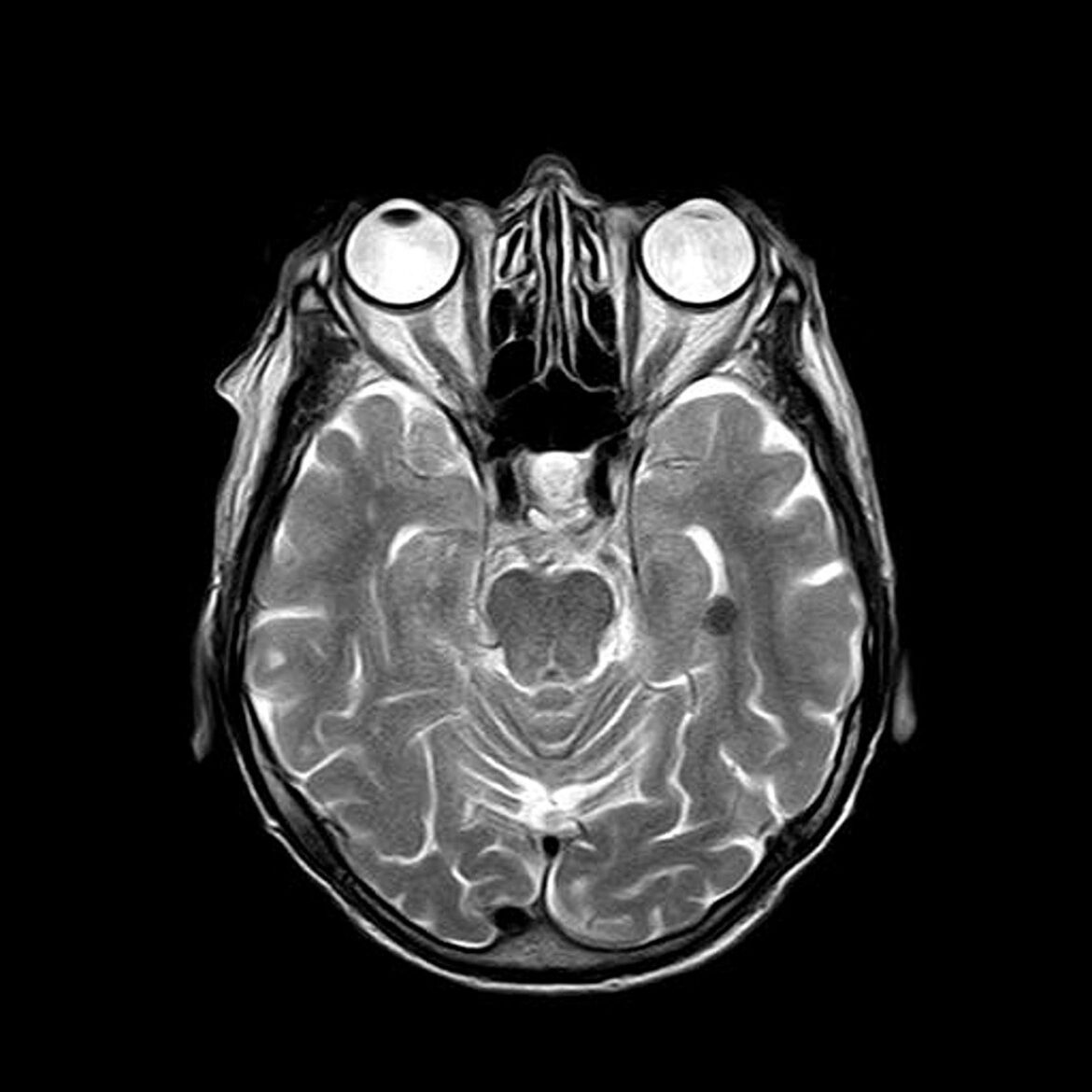
Astrocytes’ involvement in the evolution of post-traumatic epileptogenesis (PTE) has undergone a significant shift in perspective, now recognized for their complex roles that surpass their previously believed function as just supportive cells. Current studies highlight their comprehensive contributions, especially in the pathogenic inflammatory responses and their active participation in critical brain functions, including learning, memory, and sleep regulation. Astrocytes, as the brain’s most abundant cell type, are crucial in ensuring ionic balance, blood-brain barrier integrity, neurotransmitter regulation, and neuronal energy provision. They are instrumental in modulating neuronal activity, such as enhancing the exchange between neuronal pyruvate and astrocytic lactate, thus promoting neuronal metabolism and aiding in synaptic information processing by adjusting neurotransmitter uptake and release. Their regulation of neurotransmitters like glutamate and GABA significantly affects synaptic communication.
Within the PTE framework, astrocyte activation forms a core part of the neuroinflammatory reaction. Following head injury, gliosis, a marker of astrocyte activation, is frequently detected. This condition, known as astrocytosis, occurs both at the injury site and in adjacent brain regions, posing challenges in distinguishing between gliosis induced by seizures and that which may predispose to epilepsy. Yet, similar patterns of gliosis have been identified across various animal models of traumatic brain injury (TBI), providing insights into astrocytic roles in epileptogenesis.
Post-TBI, astrocytes respond to axonal damage, neuronal death, and the rapid release of inflammatory molecules, affecting their physiological functions related to signaling and epileptogenesis. Research by Steinhauser and colleagues identified functional changes in astrocytes in epileptic conditions, such as decreased potassium currents and altered gap junction connectivity, essential in the development of epilepsy. Astrocyte activation also elevates intracellular calcium levels, increasing glutamate release, which may lead to neuronal excitotoxicity and heightened seizure susceptibility, potentially through inflammation-induced receptor expression changes and gap junction disruptions.
The disconnection of astrocyte gap junctions is a notable aspect of their activation following head trauma. Additionally, the involvement of astrocytic Cx43 hemichannels in seizures has been documented, with interventions like GAP19, a hemichannel inhibitor, showing potential in reducing seizure activities, suggesting that neuroinflammatory responses to TBI might impair astrocytes’ ability to manage ion balance effectively.
Neuroinflammation also prompts both functional and structural changes in astrocytes, linked to seizure and epilepsy risks. For example, alterations in aquaporin-4 function and distribution are thought to increase susceptibility to post-traumatic seizures (PTS), while astrocyte hypertrophy following head trauma could facilitate the development of pro-epileptogenic circuits.
The role of microglia and cytokines post-TBI is also critical, with microglial activation persisting long after the initial injury. These cells, central to the CNS’s immune defense, contribute to neuronal proliferation, differentiation, and synaptic connectivity. Microglial activation, characteristic of both TBI and epileptogenesis, involves migrating to injury sites, isolating damaged tissues, and phagocytizing debris. Inhibiting microglial activity has been shown to mitigate PTS, cognitive deficits, and epilepsy, underscoring their role in neuronal hyperexcitability. The activation process is initiated by chemokines and pro-inflammatory cytokines, with the DAMP protein HMGB1, for instance, playing a significant role in triggering seizure-resistant pathways.
Microglia can exhibit both beneficial and detrimental effects on PTE development, with evidence pointing to different activation states (M1 and M2) that may influence the acute response and long-term neurological outcomes. The interaction between microglia and astrocytes is crucial in PTE neuroinflammation, where activated microglia can induce a neurotoxic A1 astrocyte phenotype, contributing to neuronal damage and synaptic disassembly.
Cytokines and chemokines significantly impact PTE, with their rapid and sustained release after TBI documented. Elevated levels of specific cytokines, such as IL-1β, TGFβ, TNF, IL-6, and IL-10, after TBI, directly and indirectly heighten neuronal excitability and seizure susceptibility, highlighting their critical roles in the pathophysiology of post-traumatic epilepsy.
| Feature | Description |
|---|---|
| Astrocytes’ Role Beyond Support | Traditionally considered support cells, astrocytes are now recognized for their multifaceted roles in brain functions including learning, memory, and sleep, and in the development of post-traumatic epileptogenesis (PTE). |
| Maintenance Functions | Astrocytes are pivotal in maintaining ionic homeostasis, preserving blood-brain barrier integrity, regulating neurotransmitter metabolism, and ensuring neuronal energy supply. |
| Modulation of Neuronal Activity | They facilitate neuronal metabolism by exchanging neuronal pyruvate for astrocytic lactate and participate in synaptic information processing through modulation of neurotransmitter uptake and release. |
| Neuroinflammatory Response to TBI | Astrocyte activation is a significant part of the neuroinflammatory response to traumatic brain injury (TBI), contributing to the pathophysiology of PTE. |
| Response to Axonal Degeneration and Cell Death | In response to TBI, astrocytes react to axonal degeneration and neuronal cell death, which can alter their physiological functions and impact epileptogenesis. |
| Functional Changes in Epileptic Conditions | Research has identified functional changes in astrocytes in epileptic brains, including reduced potassium currents and altered gap junction coupling, which are crucial in epilepsy development. |
Literature – further reading:
- Kettenmann, H.; Verkhratsky, A.Neuroglia: The 150 years after. Trends Neurosci. 2008, 31, 653–659.
- Verkhratsky, A.; Ho, M.S.; Vardjan, N.; Zorec, R.; Parpura, V. General pathophysiology of astroglia. In Neuroglia in Neurodegenerative Diseases; Advances in Experimental Medicine and Biology; Springer : Singapore, 2019; Volume 1175, pp. 149–179.
- Oberheim, N.A.; Takano, T.; Han, X.; He, W.; Lin, J.H.C.; Wang, F.; Xu, Q.; Wyatt, J.D.; Pilcher, W.; Ojemann, J.; et al. Uniquely Hominid Features of Adult Human Astrocytes. J. Neurosci. 2009, 29, 3276–3287.
- Halassa, M.M.; Haydon, P.G. Integrated brain circuits: Astrocytic networks modulate neuronal activity and behavior. Annu. Rev. Physiol. 2010, 72, 335–355.
- Araque, A.; Carmignoto, G.; Haydon, P.G.; Oliet, S.H.; Robitaille, R.; Volterra, A. Gliotransmitters travel in time and space.
Neuron 2014, 81, 728–739. - Weber, B.; Barros, L.F. The Astrocyte: Powerhouse and Recycling Center. Cold Spring Harb. Perspect. Biol. 2015, 7, 12.
- Ye, Z.-C.; Sontheimer, H. Cytokine modulation of glial glutamate uptake: A possible involvement of nitric oxide. Neuroreport 1996, 7, 2181–2185.
- Zhu, G.; Okada, M.; Yoshida, S.; Mori, F.; Ueno, S.; Wakabayashi, K.; Kaneko, S. Effects of interleukin-1beta on hippocampal glutamate and GABA releases associated with Ca2+-induced Ca2+ releasing systems. Epilepsy Res. 2006, 71, 107–116.
- Hu, S.; Sheng, W.S.; Ehrlich, L.C.; Peterson, P.K.; Chao, C.C. Cytokine effects on glutamate uptake by human astrocytes. Neuroimmunomodulation 2000, 7, 153–159.
- Payan, H.; Toga, M.; Bérard-Badier, M. The pathology of post-traumatic epilepsies. Epilepsia 1970, 11, 81–94.
- Oehmichen, M.; Walter, T.; Meissner, C.; Friedrich, H.J. Time course of cortical hemorrhages after closed traumatic brain injury: Statistical analysis of posttraumatic histomorphological alterations. J. Neurotrauma 2003, 20, 87–103.
- Pelinka, L.E.; Kroepfl, A.; Leixnering, M.; Buchinger, W.; Raabe, A.; Redl, H. GFAP versus S100B in serum after traumatic brain injury: Relationship to brain damage and outcome. J. Neurotrauma 2004, 21, 1553–1561.
- Swartz, B.E.; Houser, C.R.; Tomiyasu, U.; Walsh, G.O.; DeSalles, A.; Rich, J.R.; Delgado-Escueta, A. Hippocampal cell loss in
posttraumatic human epilepsy. Epilepsia 2006, 47, 1373–1382. - Van Landeghem, F.K.H.; Weiss, T.; Oehmichen, M.; Deimling, A. Von Decreased Expression of Glutamate Transporters in Astrocytes after Human Traumatic Brain Injury. J. Neurotrauma 2006, 23, 1518–1528.
- Brooks, D.M.; Patel, S.A.; Wohlgehagen, E.D.; Semmens, E.O.; Pearce, A.; Sorich, E.A.; Rau, T.F. Multiple mild traumatic brain injury in the rat produces persistent pathological alterations in the brain. Exp. Neurol. 2017, 297, 62–72.
- Domowicz, M.; Wadlington, N.L.; Henry, J.G.; Diaz, K.; Munoz, M.J.; Schwartz, N.B. Glial cell responses in a murine multifactorial perinatal brain injury model. Brain Res. J. 2018, 1681, 52–63.
- Bye, N.; Carron, S.; Han, X.; Agyapomaa, D.; Ng, S.Y.; Yan, E.; Rosenfeld, J.V.; Morganti-Kossmann, M.C. Neurogenesis and
glial proliferation are stimulated following diffuse traumatic brain injury in adult rats. J. Neurosci. Res. 2011, 89, 986–1000.
Budde, M.D.; Janes, L.; Gold, E.; Turtzo, L.C.; Frank, J.A. The contribution of gliosis to diffusion tensor anisotropy and tractography following traumatic brain injury: Validation in the rat using Fourier analysis of stained tissue sections. Brain 2011, 134,2248–2260.

